Copyright
©The Author(s) 2021.
World J Clin Cases. Aug 26, 2021; 9(24): 7231-7236
Published online Aug 26, 2021. doi: 10.12998/wjcc.v9.i24.7231
Published online Aug 26, 2021. doi: 10.12998/wjcc.v9.i24.7231
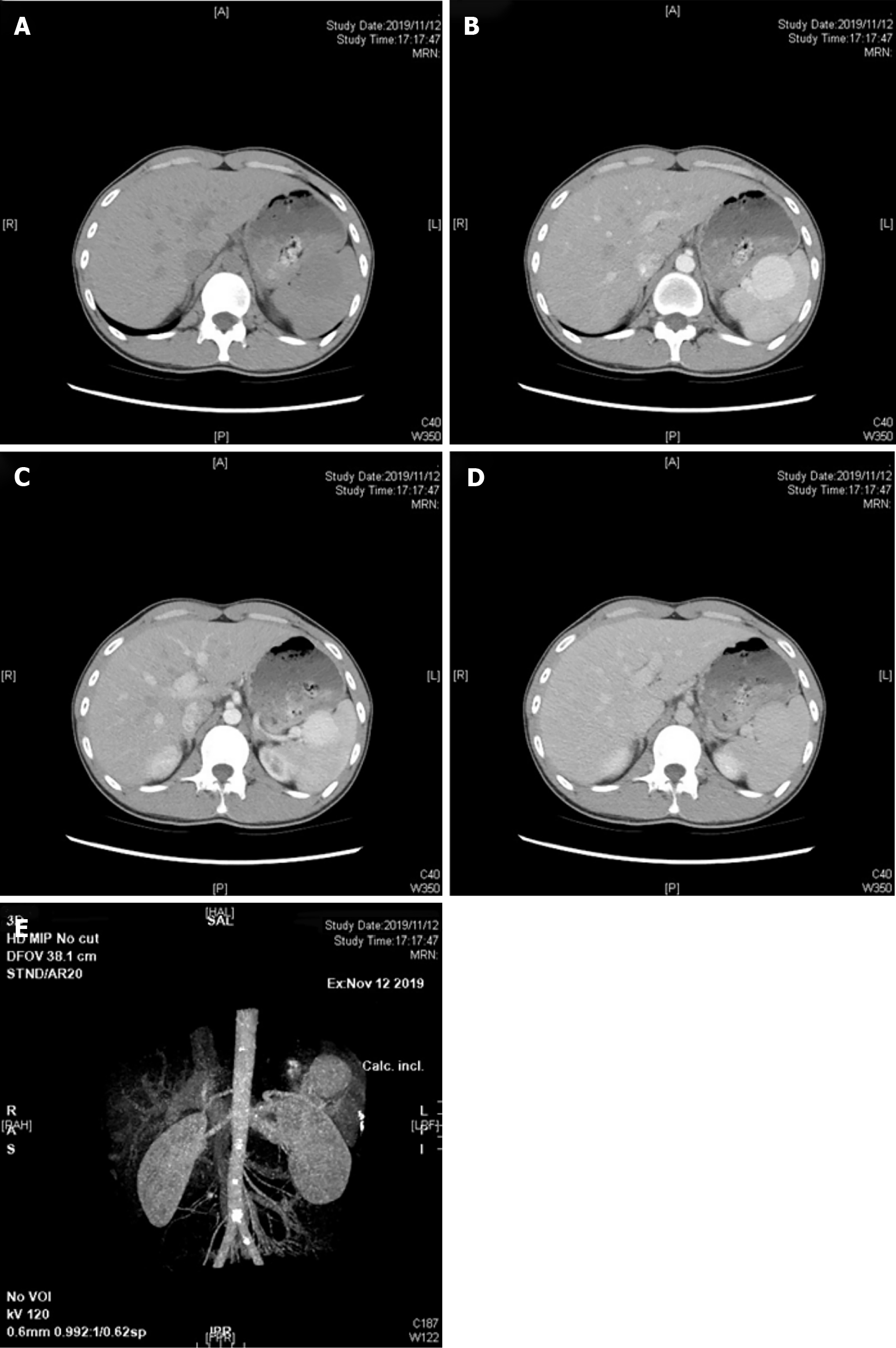
Figure 1 The manifestation of tumor in different computed tomography phases.
A: Computed tomography (CT) findings in plain phase; B: CT findings in the arterial phase; C: CT findings in the venous phase; D: CT findings in the delayed phase; E: CT findings of three-dimensional reconstruction of the artery.
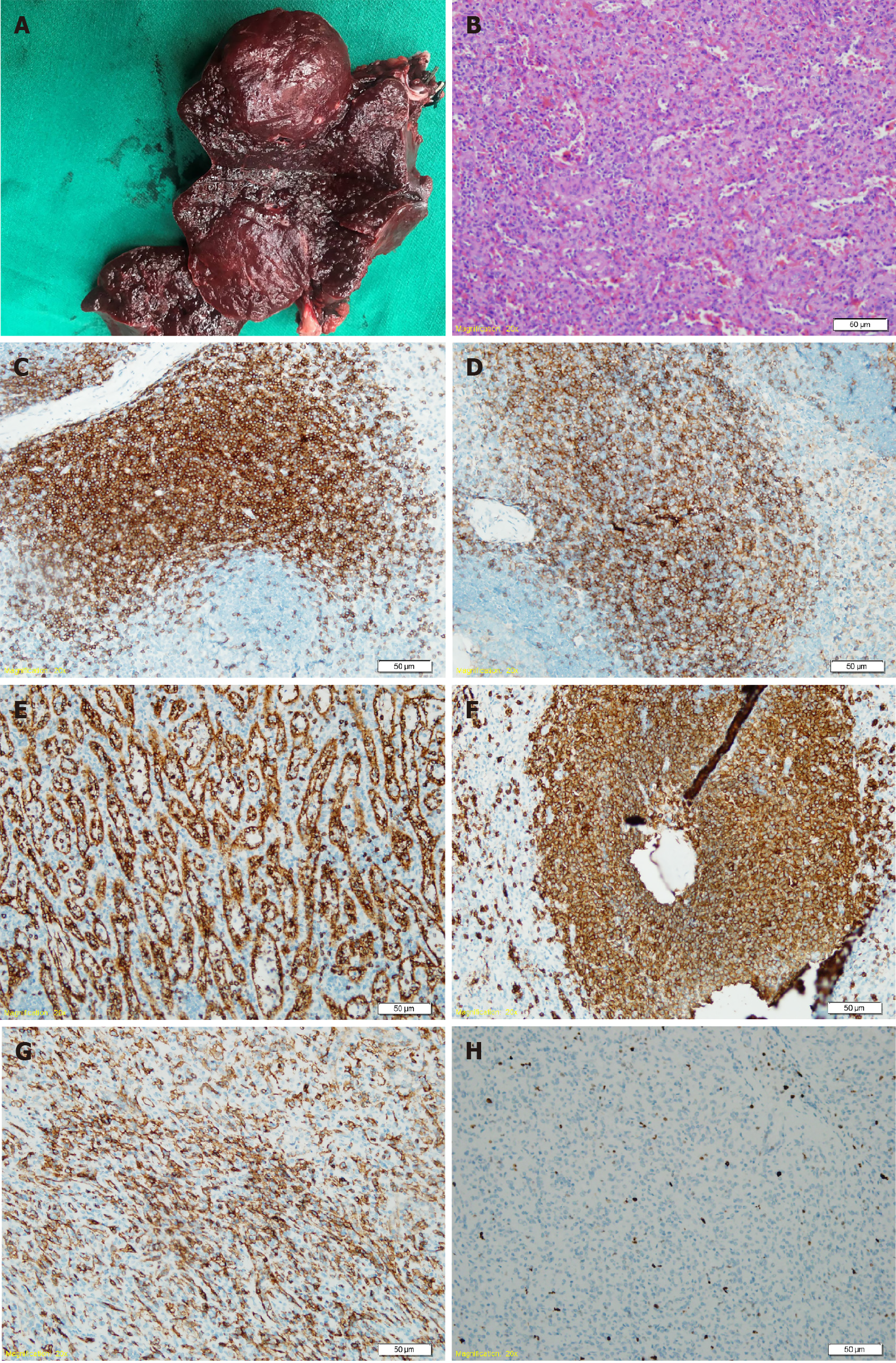
Figure 2 The postoperative gross specimen and pathological examination.
A: Postoperative gross specimen; B: Hematoxylin and eosin staining of the lesion; C: Cluster of differentiation 3 (CD3) staining in immunohistochemistry (IHC); D: CD4 staining in IHC; E: CD8 staining in IHC; F: CD20 staining in IHC; G: CD34 staining in IHC; H: Ki-67 staining in IHC.
- Citation: Cao XF, Yang LP, Fan SS, Wei Q, Lin XT, Zhang XY, Kong LQ. Incidentally discovered asymptomatic splenic hamartoma misdiagnosed as an aneurysm: A case report. World J Clin Cases 2021; 9(24): 7231-7236
- URL: https://www.wjgnet.com/2307-8960/full/v9/i24/7231.htm
- DOI: https://dx.doi.org/10.12998/wjcc.v9.i24.7231


