Copyright
©The Author(s) 2021.
World J Clin Cases. Aug 26, 2021; 9(24): 7212-7217
Published online Aug 26, 2021. doi: 10.12998/wjcc.v9.i24.7212
Published online Aug 26, 2021. doi: 10.12998/wjcc.v9.i24.7212
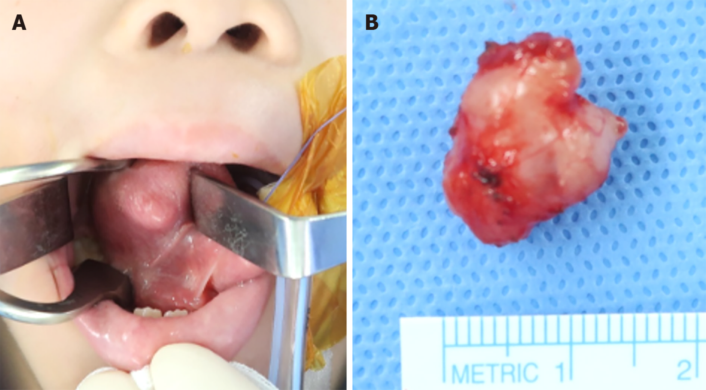
Figure 1 Intraoperative view.
A: Preoperative view of the tongue lesion with intact ventral mucosa; B: Macroscopic view of the well-encapsulated, 17 mm × 14 mm × 7 mm sized tumor.
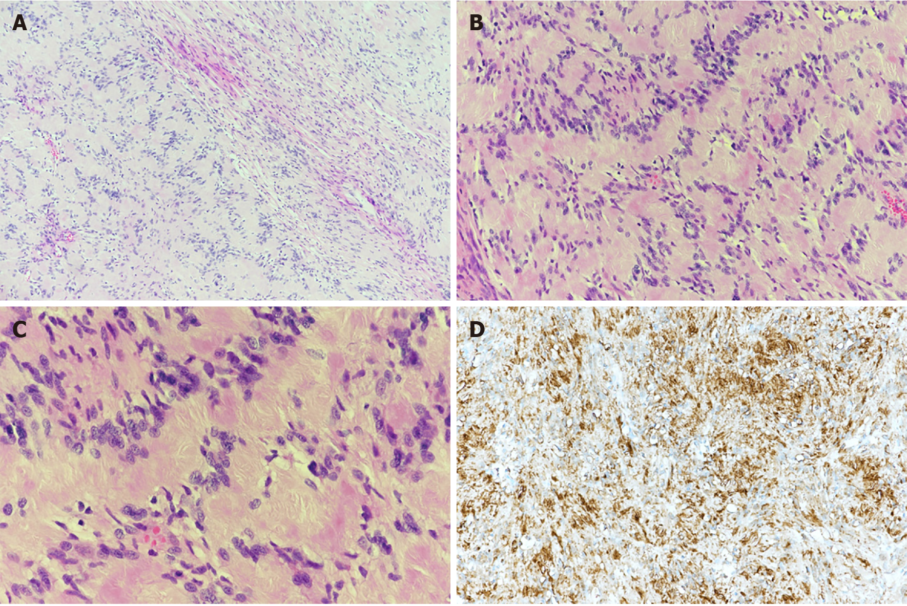
Figure 2 Hematoxylin and eosin stained.
A: Histological picture showing the typical biphasic appearance of schwannoma [hematoxylin and eosin (H&E) stained, original magnification × 100]. Densely packed spindle cells (Antoni A areas, left side) with a typical palisading arrangement (Verocay bodies) to loose hypocellular arrangements (Antoni B areas, right side); B and C: Antoni A areas composed of Verocay bodies which consists of a stacked arrangement of two rows of elongated palisading nuclei that alternates with acellular zones (H&E, × 200 and × 400); D: Immunohistochemical staining with S-100 (× 200) was strong and diffusely positive.
- Citation: Yun CB, Kim YM, Choi JS, Kim JW. Pediatric schwannoma of the tongue: A case report and review of literature. World J Clin Cases 2021; 9(24): 7212-7217
- URL: https://www.wjgnet.com/2307-8960/full/v9/i24/7212.htm
- DOI: https://dx.doi.org/10.12998/wjcc.v9.i24.7212


