Copyright
©The Author(s) 2021.
World J Clin Cases. Aug 26, 2021; 9(24): 7163-7168
Published online Aug 26, 2021. doi: 10.12998/wjcc.v9.i24.7163
Published online Aug 26, 2021. doi: 10.12998/wjcc.v9.i24.7163
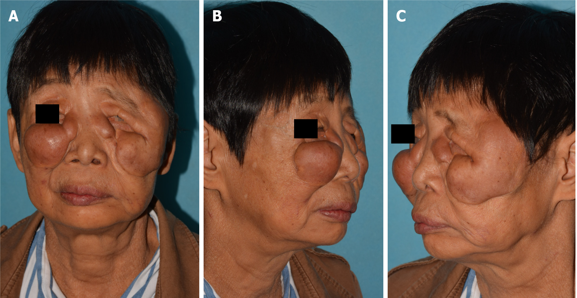
Figure 1 Clinical images of the patient.
A: Front view; B and C: Lateral view. The masses were distributed among the upper two thirds of the patient’s face and caused severe facial disfigurement and pupillary axis obstruction of the left eye.
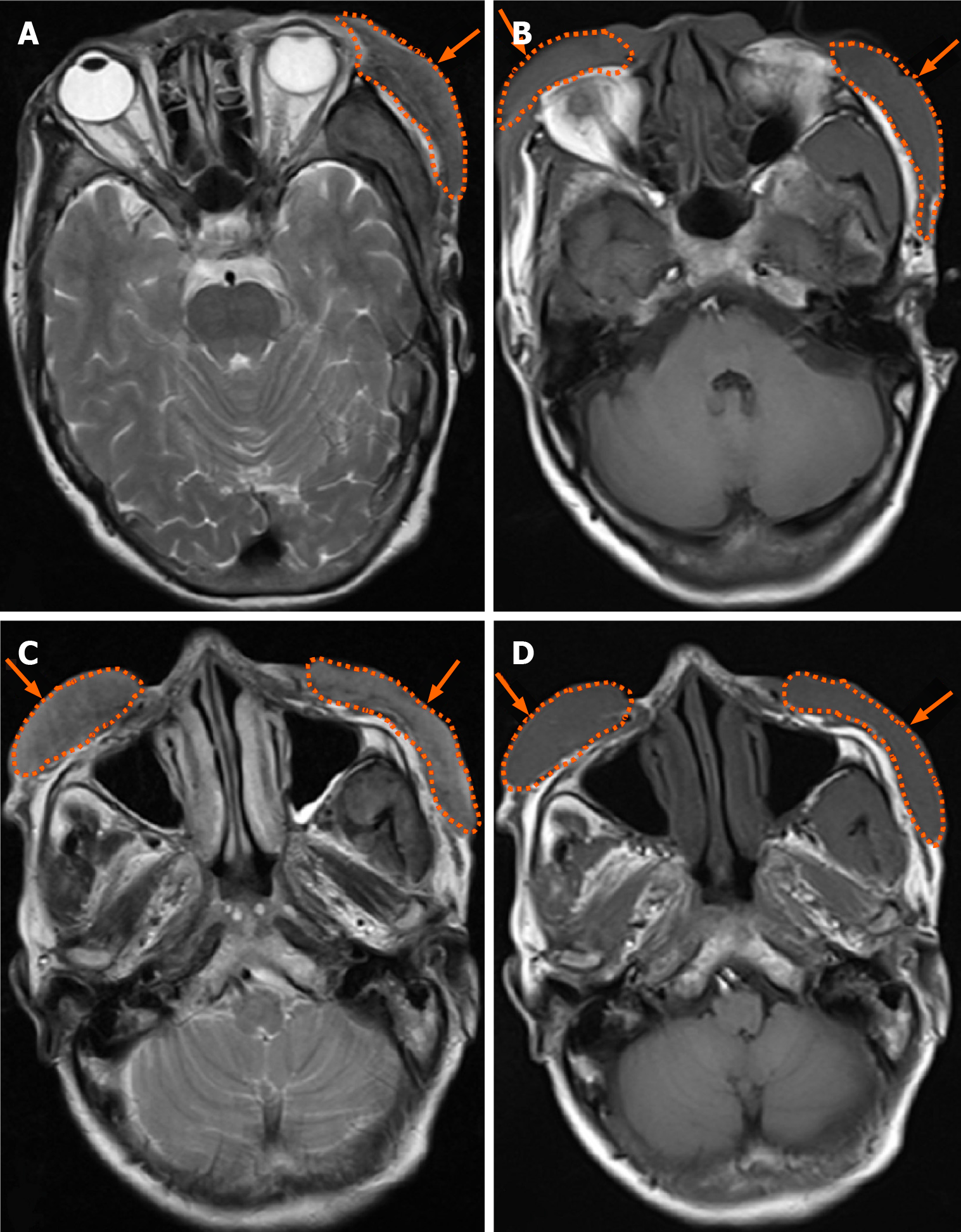
Figure 2 Imaging examination.
Axial magnetic resonance imaging of the patient’s head showed bilateral periorbital and left temporal soft tissue swelling (orange arrow) without cranium and intra-orbital invasion, and T1 weighted imaging (T1WI) and T2 weighted imaging (T2WI) showed iso-density signals. A: Left temporal soft tissue swelling on T2WI; B: Bilateral periorbital soft tissue swelling on T1WI; C: Bilateral infraorbital and zygomatic facial swelling on T2WI; D: Bilateral infraorbital and zygomatic facial swelling on T1WI.
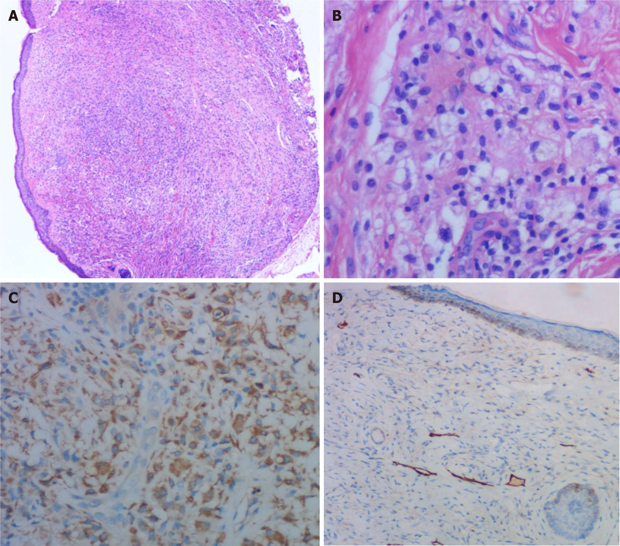
Figure 3 Histological and immunohistochemistry images.
A: Large amount of lymphocyte and histiocyte infiltration in clear-cut granulomas (hematoxylin-eosin staining, × 40); B: The infiltration of lymphocytes, foam-like histiocytes, multinucleated giant cells, and mast cells (hematoxylin-eosin staining, × 200); C: Positive expression of CD68 in histiocytes in clear-cut granulomas (immunohistochemical staining, × 100); D: Positive expression of D2-40 in the lymphatic endothelium and lymphatic vessel dilatation (immunohistochemical staining, × 100).
- Citation: Zhang L, Yan S, Pan L, Wu SF. Progressive disfiguring facial masses with pupillary axis obstruction from Morbihan syndrome: A case report. World J Clin Cases 2021; 9(24): 7163-7168
- URL: https://www.wjgnet.com/2307-8960/full/v9/i24/7163.htm
- DOI: https://dx.doi.org/10.12998/wjcc.v9.i24.7163


