Copyright
©The Author(s) 2021.
World J Clin Cases. Aug 26, 2021; 9(24): 7146-7153
Published online Aug 26, 2021. doi: 10.12998/wjcc.v9.i24.7146
Published online Aug 26, 2021. doi: 10.12998/wjcc.v9.i24.7146
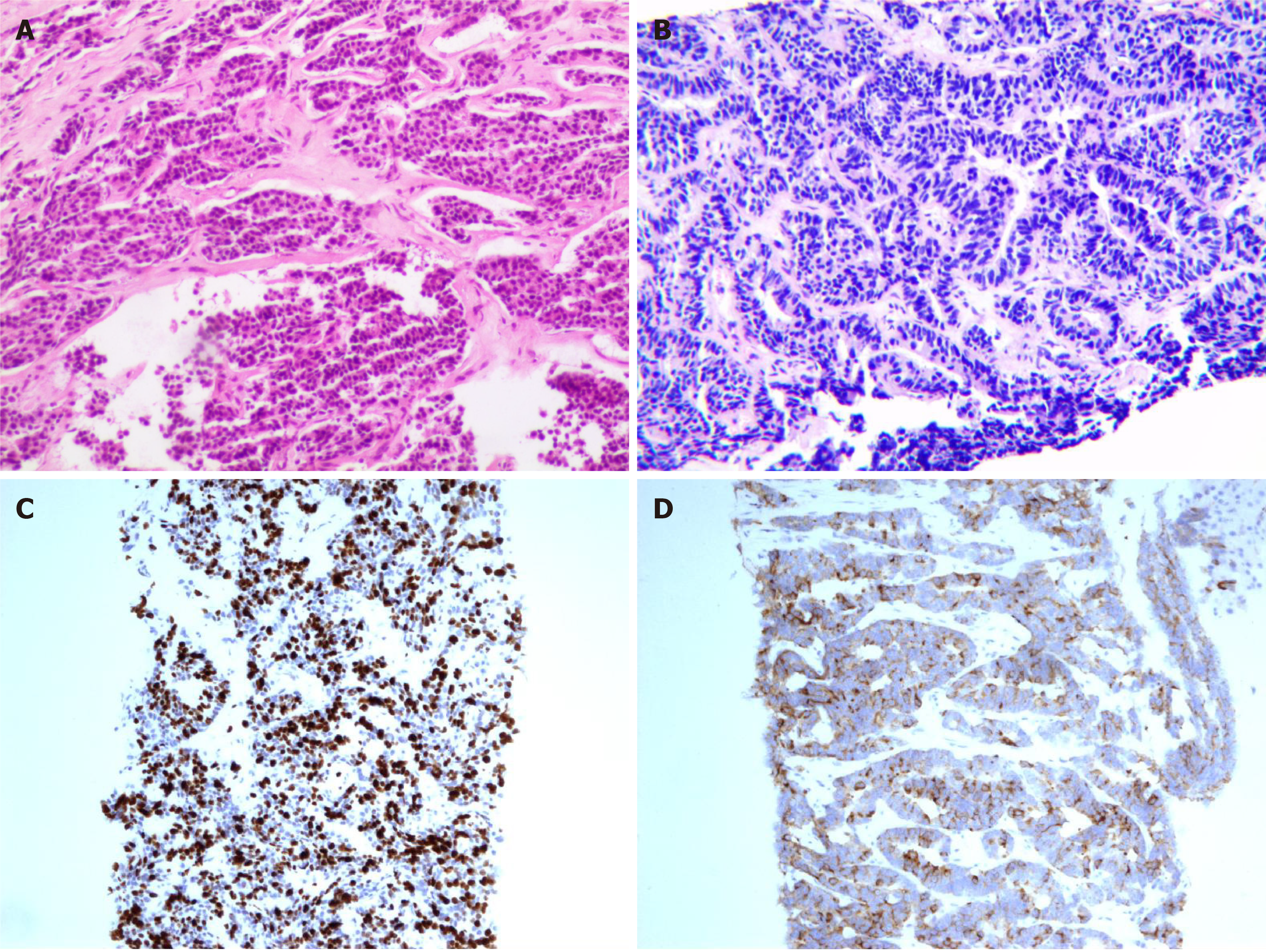
Figure 1 Pathological analysis and immunohistochemical staining.
A: Hematoxylin and eosin (100 ×) staining of right breast tissue; B: Hematoxylin and eosin (100 ×) staining of liver core biopsy specimen; C: Ki-67 index of 70%; D: Immunohistochemical staining (100 ×) reveals positivity for synaptophysin.
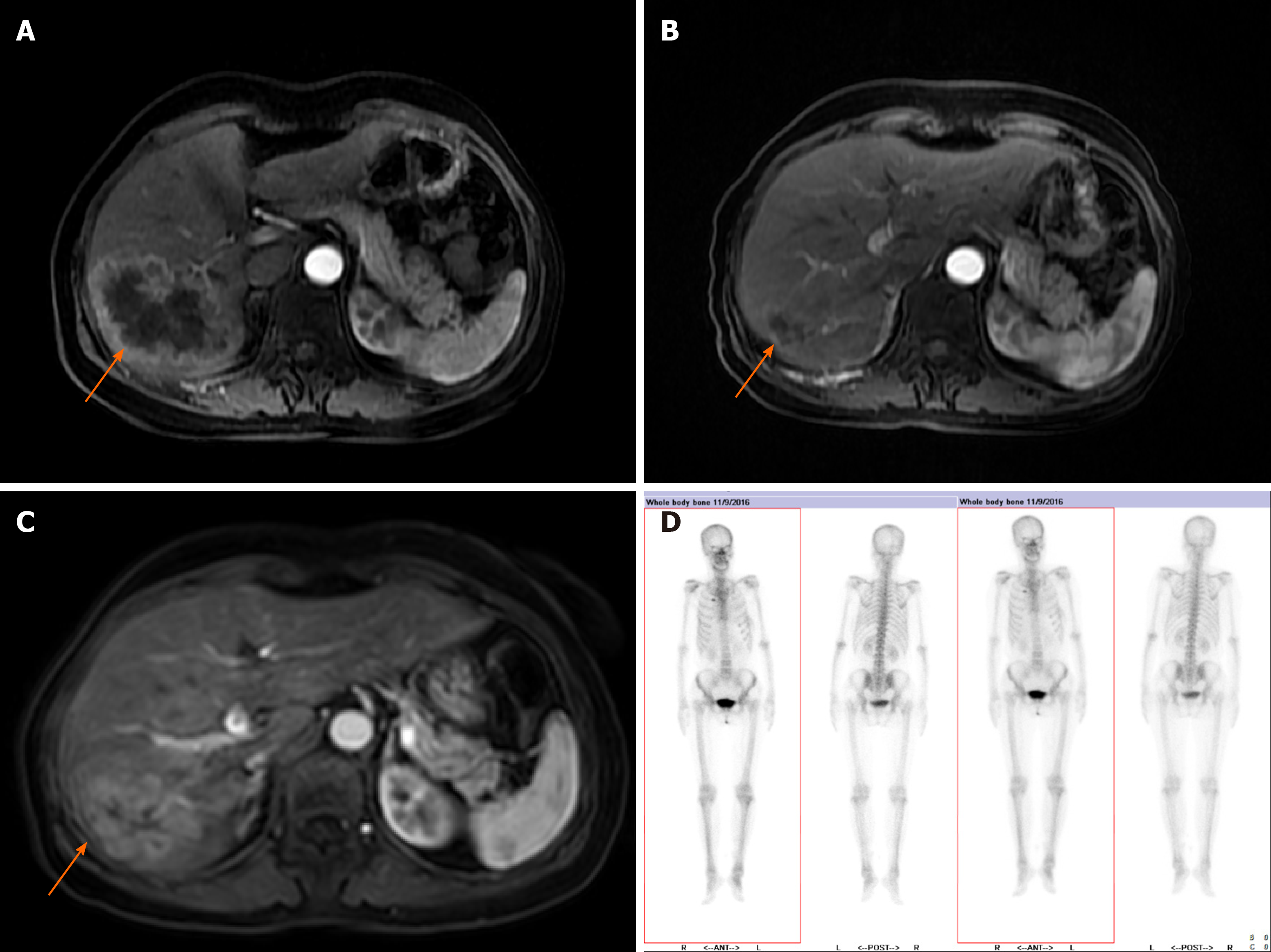
Figure 2 Follow-up abdominal magnetic resonance imaging at the beginning, 1 year, and 2 years post administration of STEM chemotherapy.
A: Abdominal magnetic resonance imaging (MRI) of the patient with hepatic metastasis performed in October 2017; B: MRI in November 2018; C: MRI in September 2019. D: Whole-body bone scans performed in November 2016. In October 2017, the enhanced image revealed a large mass of the right lobe of the liver, with marked enhancement of the edge, which was reduced in November 2018 and had progressed by September 2019. The whole-body bone scans showed right second anterior rib metastases.
- Citation: Wang X, Shi YF, Duan JH, Wang C, Tan HY. S-1 plus temozolomide as second-line treatment for neuroendocrine carcinoma of the breast: A case report. World J Clin Cases 2021; 9(24): 7146-7153
- URL: https://www.wjgnet.com/2307-8960/full/v9/i24/7146.htm
- DOI: https://dx.doi.org/10.12998/wjcc.v9.i24.7146


