Copyright
©The Author(s) 2021.
World J Clin Cases. Aug 26, 2021; 9(24): 7133-7138
Published online Aug 26, 2021. doi: 10.12998/wjcc.v9.i24.7133
Published online Aug 26, 2021. doi: 10.12998/wjcc.v9.i24.7133
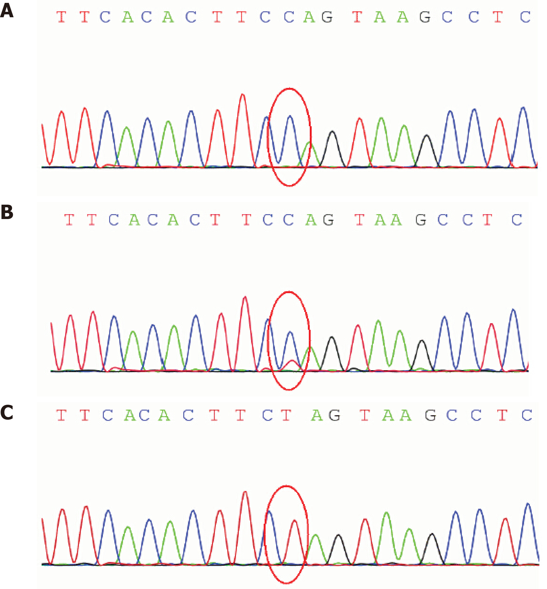
Figure 1 Sequence electropherograms showing the m.
9176T>C substitution disclosed in the patient’s and his parents’ blood samples. Restriction fragment length polymorphism (RFLP) analysis of the m.9176T>C mutation. A: The mutational load assessment by polymerase chain reaction-RFLP analysis confirmed that the patient harbored the mutation in blood leukocytes. Moreover, the variant was nearly homoplasmic (> 99%) in the tissues examined; B: The mutation was also found in maternal blood at a low level of heteroplasmy; C: The mutation was not found in DNA samples derived from the patient’s father.
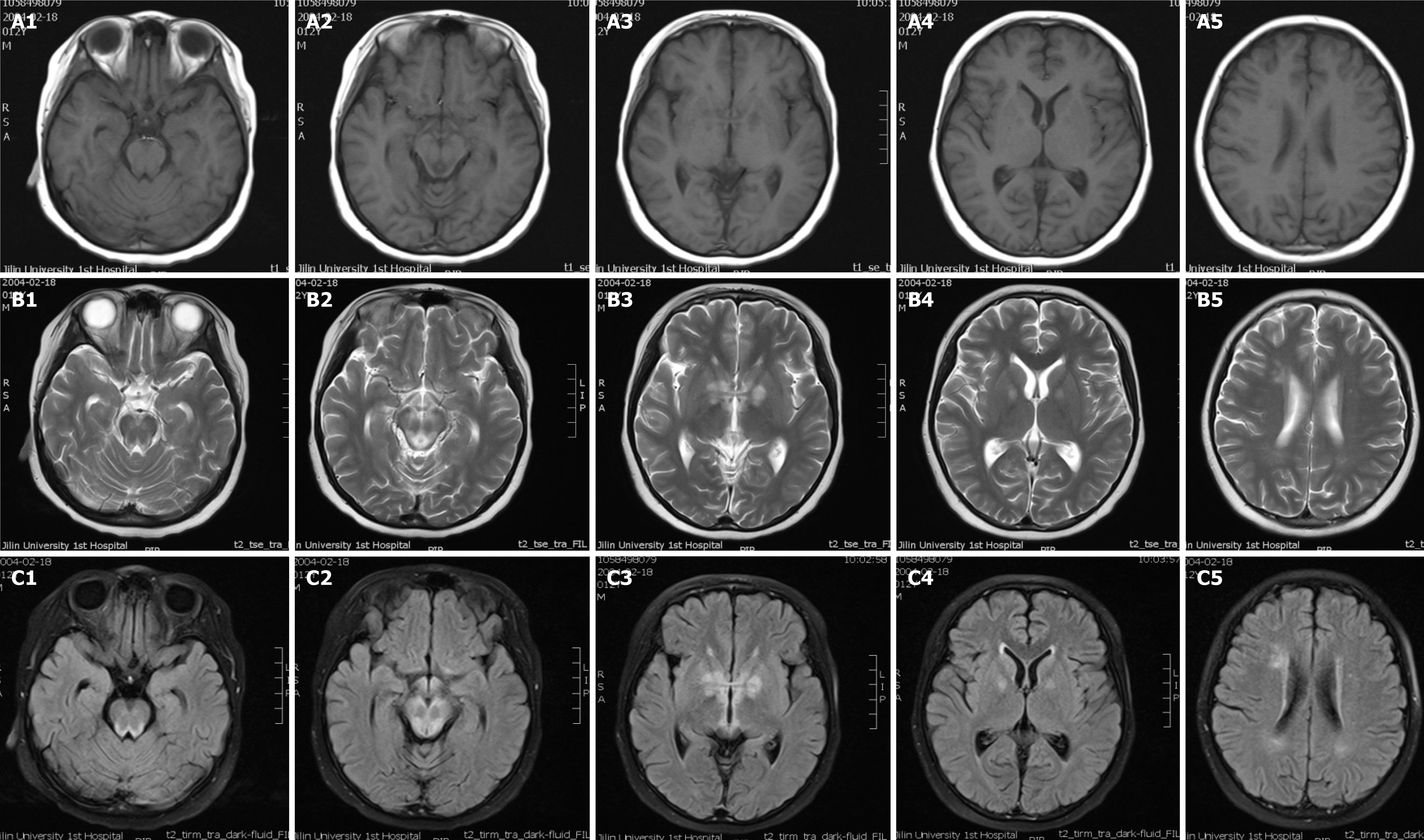
Figure 2 Neuroradiological features.
Sequential magnetic resonance imaging (MRI) scans showed bilateral symmetric signal abnormalities in the basal ganglia, medial thalami, periaqueductal region of the midbrain and pons, and bilateral white matter around the lateral ventricles (A1-5: T1-weighted images, B1-5: T2-weighted images). C1-5: Diffusion-weighted images of the brain MRI showing bilateral signal abnormalities in the basal ganglia, brain stem, and white matter.
- Citation: Liang JM, Xin CJ, Wang GL, Wu XM. Late-onset Leigh syndrome without delayed development in China: A case report. World J Clin Cases 2021; 9(24): 7133-7138
- URL: https://www.wjgnet.com/2307-8960/full/v9/i24/7133.htm
- DOI: https://dx.doi.org/10.12998/wjcc.v9.i24.7133


