Copyright
©The Author(s) 2021.
World J Clin Cases. Aug 26, 2021; 9(24): 7123-7132
Published online Aug 26, 2021. doi: 10.12998/wjcc.v9.i24.7123
Published online Aug 26, 2021. doi: 10.12998/wjcc.v9.i24.7123
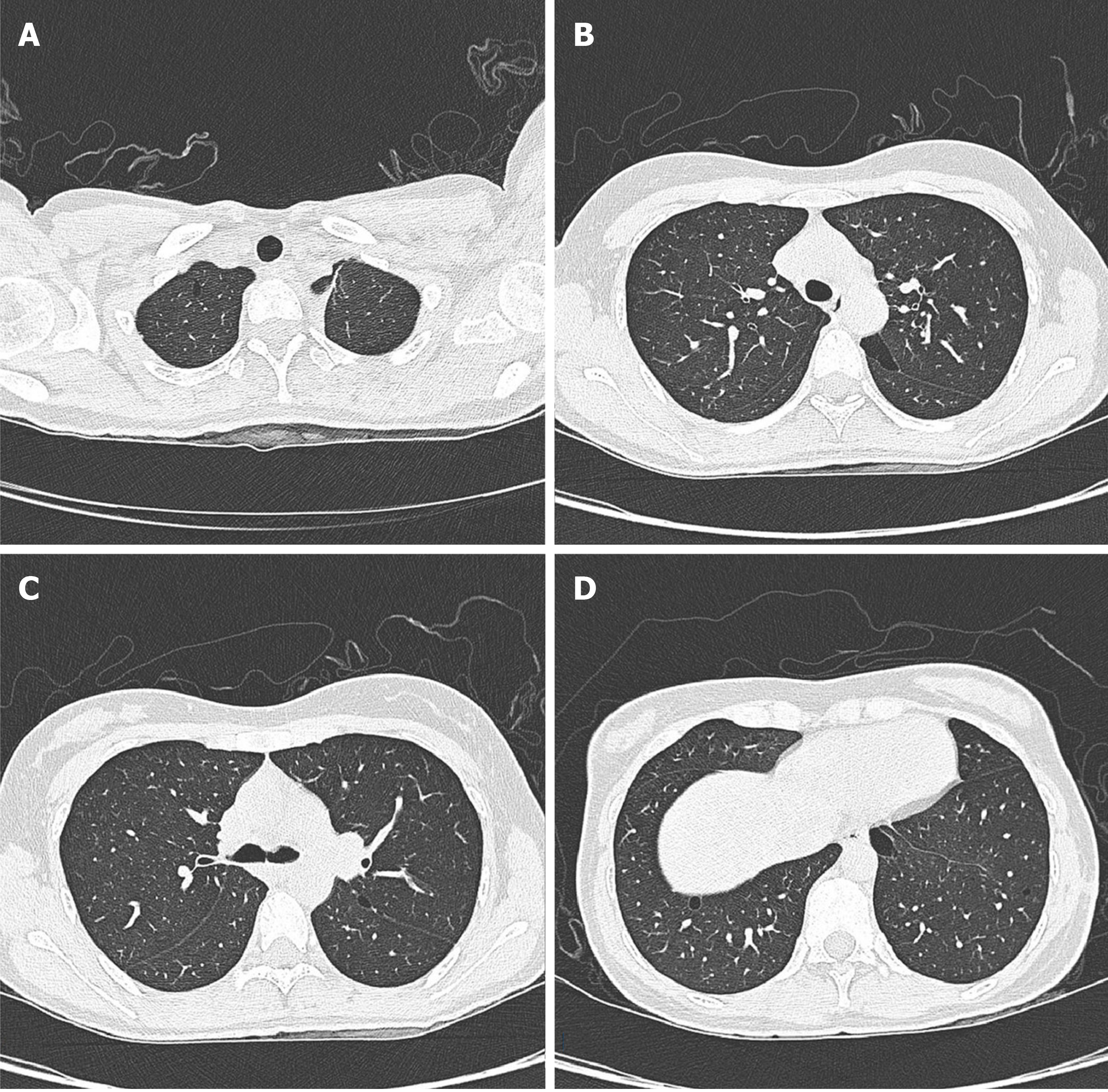
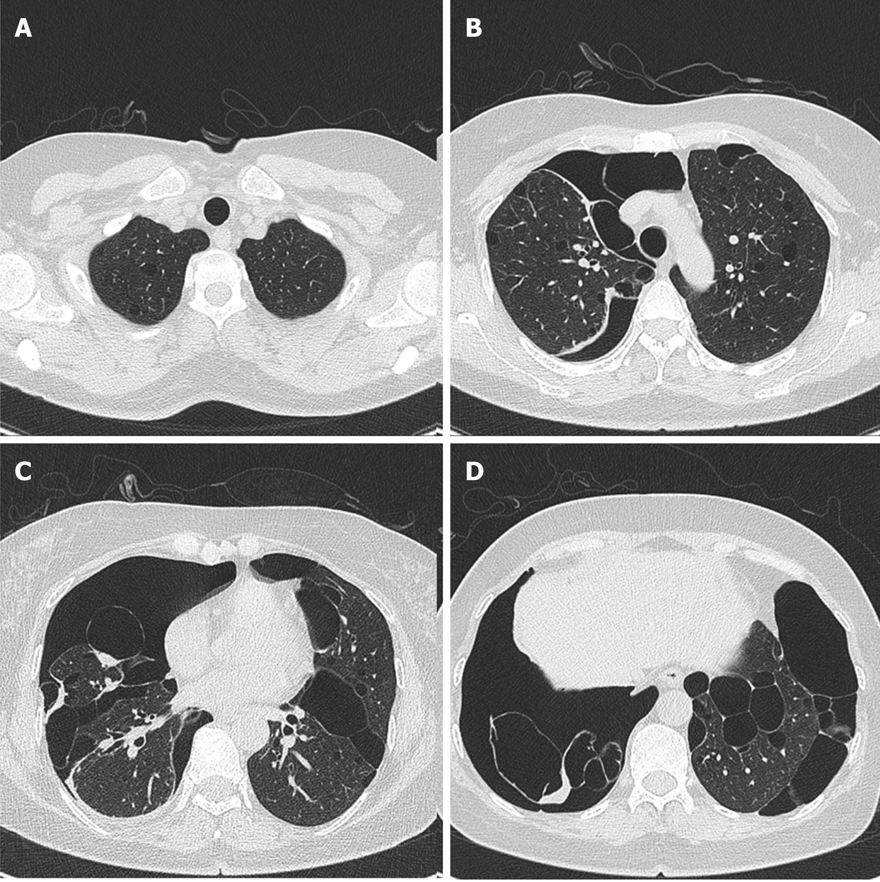
Figure 1 Patient’s axial computed tomography image showing multiple thin-walled cysts with clear margins and diverse sizes.
A: Small thin-walled cyst located in the apical segment of the right upper lobe; B and C: Irregular morphology cysts located in the apical posterior segment; D: Lower chest showing subpleural cysts at both lung bases.
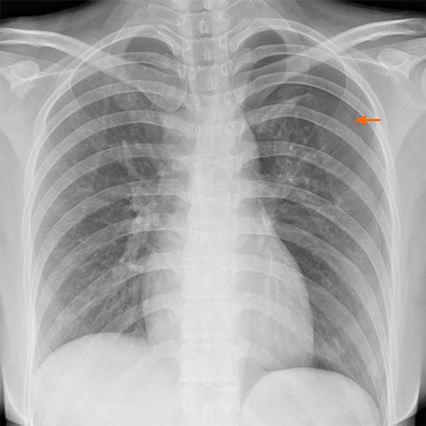
Figure 2 X-ray showing pneumothorax.
Patient’s chest X-ray showing pneumothorax in the left lung; the lung tissue was compressed to about 30%. The orange arrow represents the pneumothorax line.
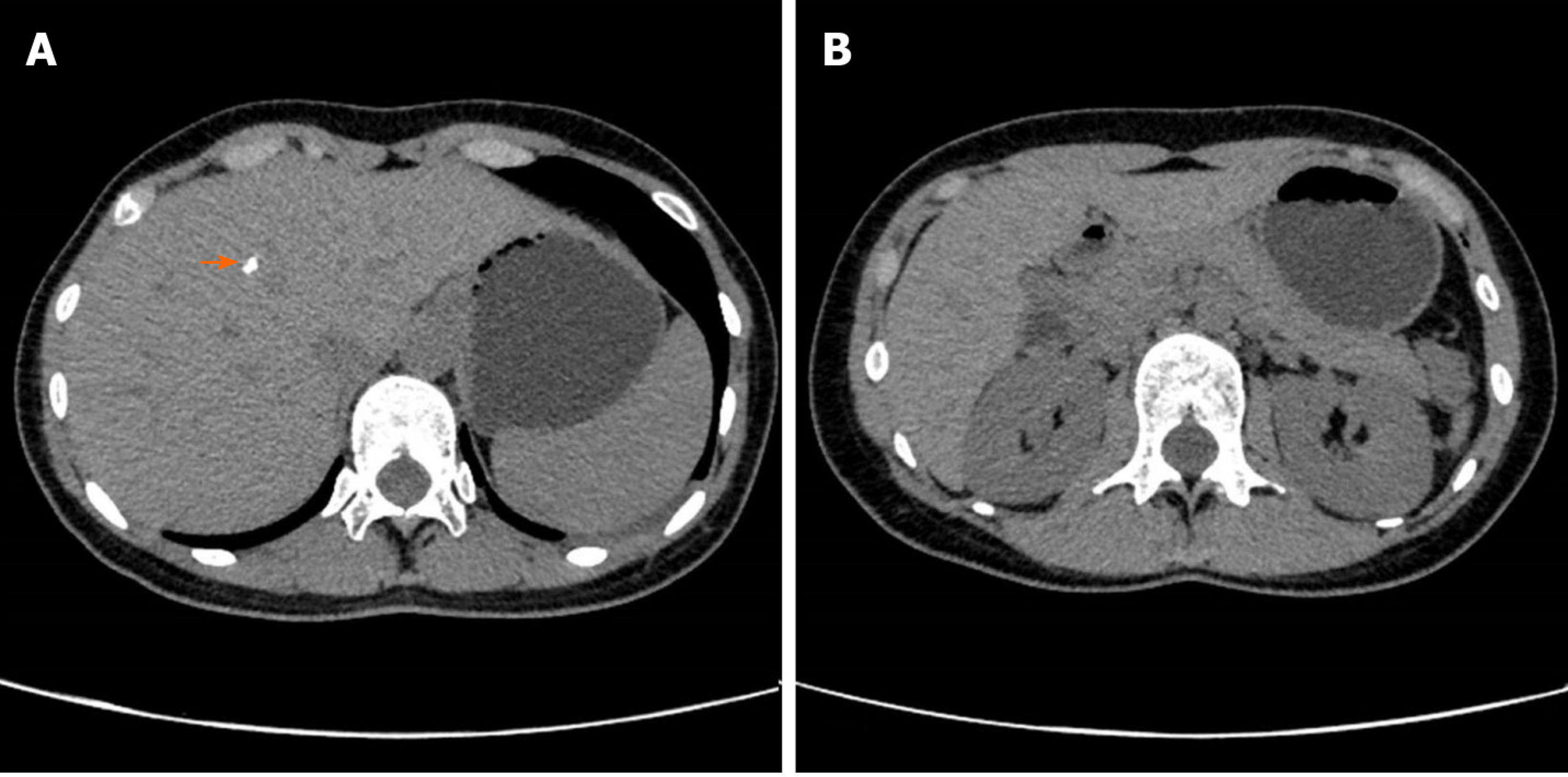
Figure 3 Abdominal computed tomography.
A: Dotted high-density shadow (orange arrow) on the right liver; B: No abnormal occupation was captured in the kidneys.
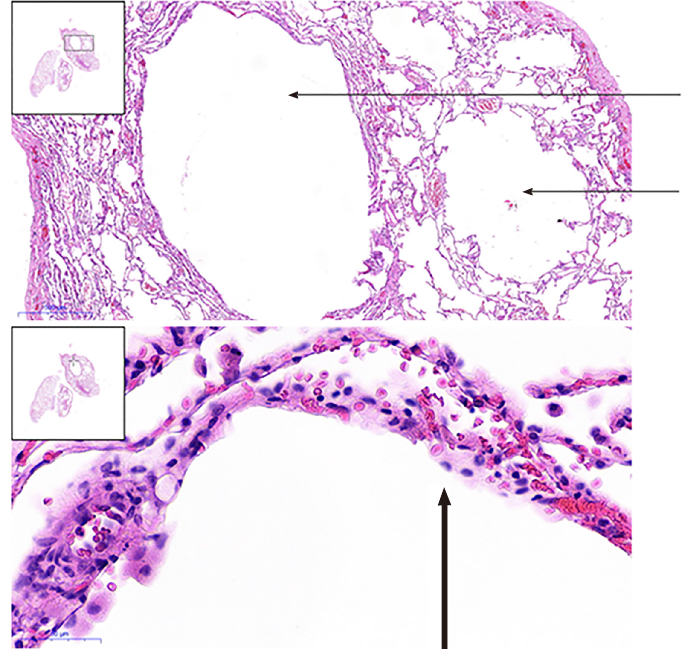
Figure 4 Postoperative pathological reports.
The postoperative pathological reports displayed the formation of multiple cysts (thin arrow). The inner surface of the pulmonary cyst was covered with alveolar epithelium (thick arrow).
Figure 5 Axial computed tomography images.
A: The patient’s mother showed multiple thin-walled cysts in the apical segment of the right upper lobe; B: Irregular morphology cysts located in the upper-lung of both sides; C: Bilateral multiple lung cysts located at the level of the upper mediastinum; D: Right-sided pneumothorax and bilateral lung cysts located at both lung bases.
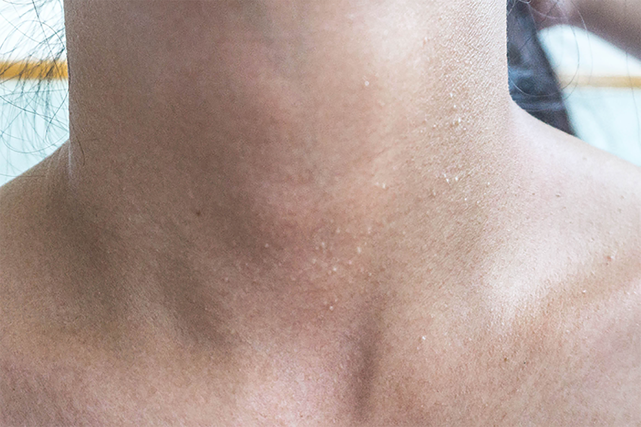
Figure 6 Multiple white dome-shaped papules visible in the neck of the patient that were consistent with the featured lesions of Birt-Hogg-Dubé syndrome.
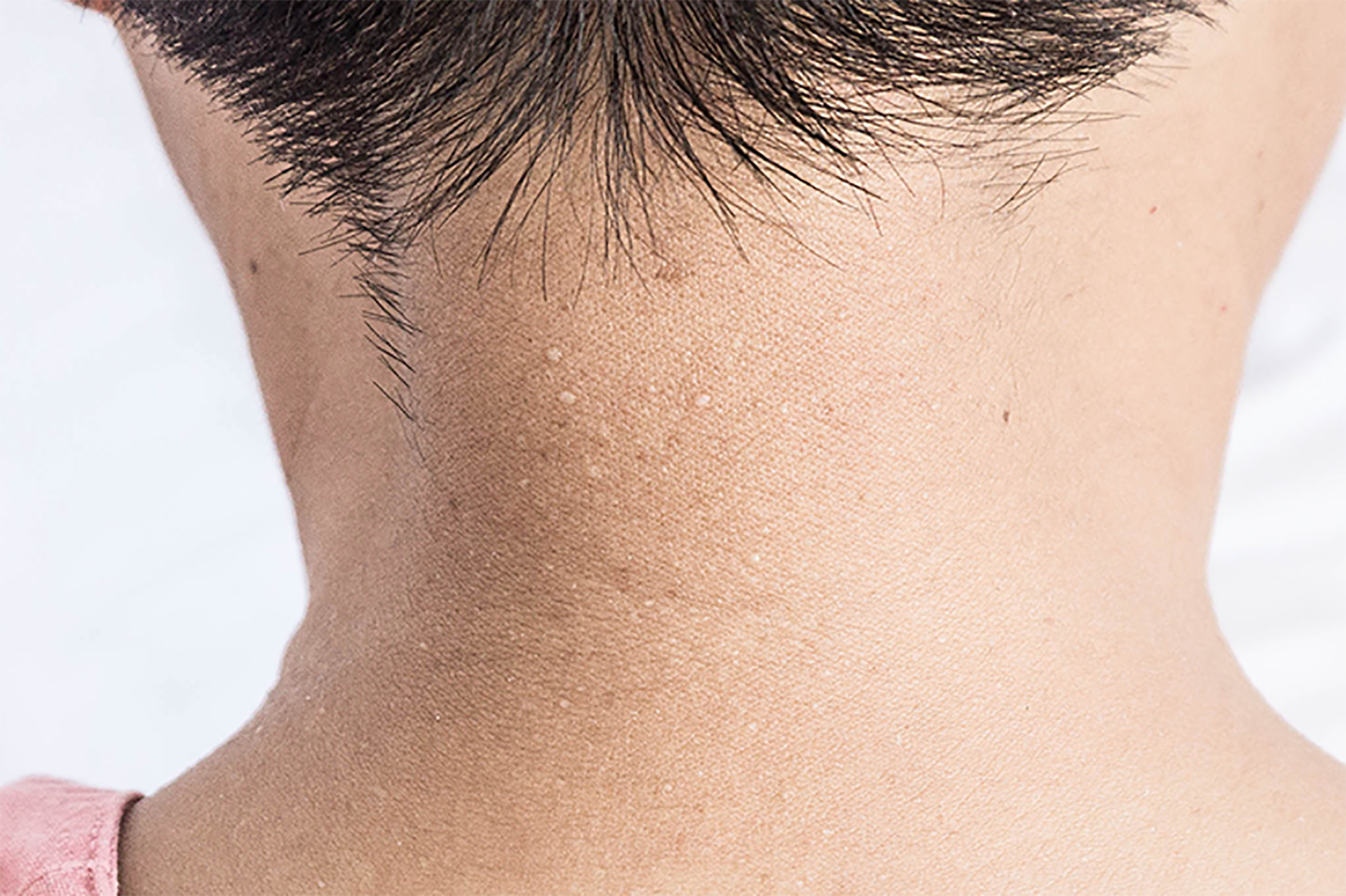
Figure 7 Multiple white dome-shaped papules were seen on the skin of her mother’s neck and back.
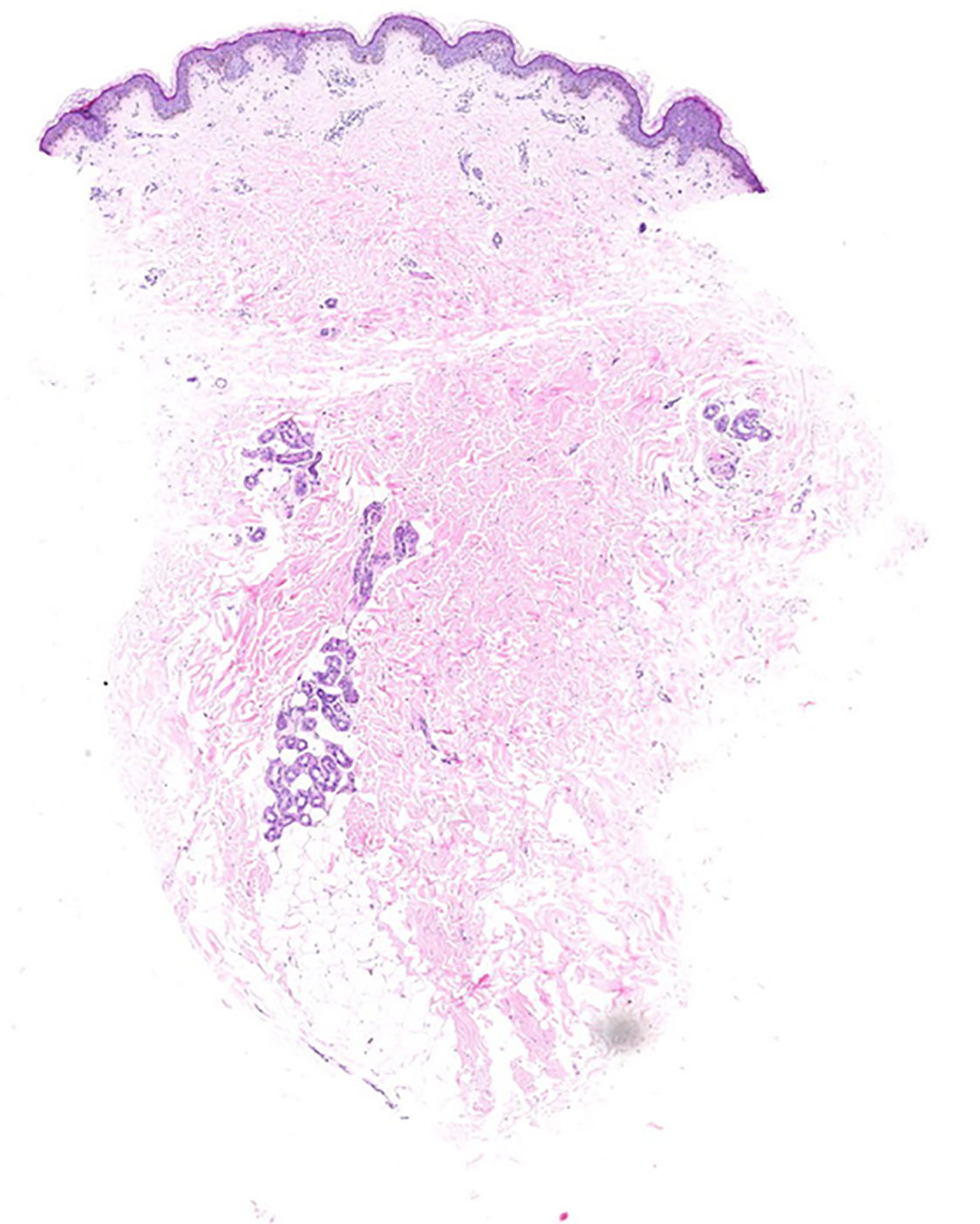
Figure 8 Pathological findings of skin biopsy showing lymphocytic infiltration around the superficial vasculature of the dermis with melanocytes.
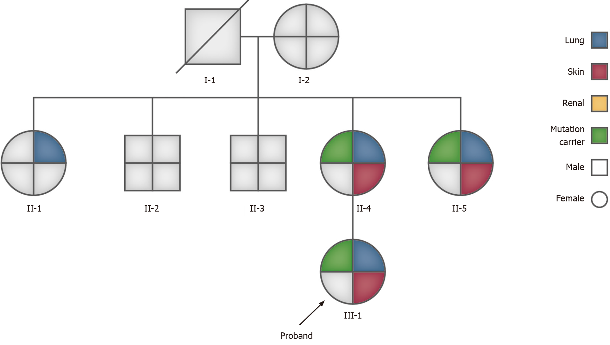
Figure 9 Seven of the patient’s family members were genetically analysed.
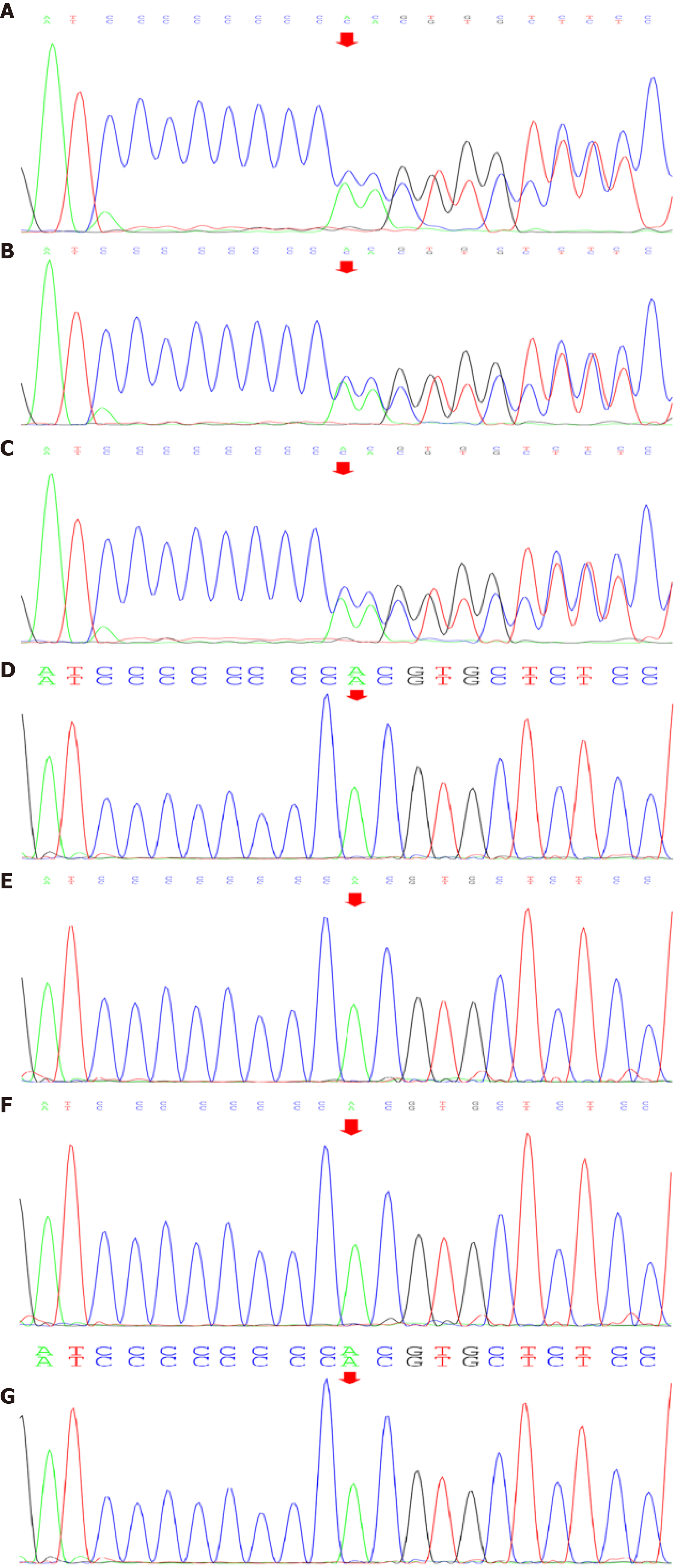
Seven of the patient’s family members were genetically analysed (I-1 was deceased), and the germline mutations in the folliculin gene mutation of the patients’ were derived from her mother. The source of her mother’s gene mutation could not be tracked due to the failure of the gene test in I-1. The patient, her mother, and her second maternal aunt had a similar genetic mutation, and the performance of their clinically affected organs was identical.
Figure 10 The American College of Medical Genetics and Genomics rating considered suggested the presence of PVS1 and PM2 by taking into consideration the suspected diseases.
A: Genetic test revealed a heterozygous nucleotide variation (red arrow) of c.1285_1286insC in germline mutations in the folliculin gene of the patient; B: The patient’s mother; C: The patient’s second maternal aunt; D: No genetic variation (red arrow) was detected in her elder aunt; E: Her elder uncle; F: Her second uncle; G: Her grandmother.
- Citation: Lu YR, Yuan Q, Liu J, Han X, Liu M, Liu QQ, Wang YG. A rare occurrence of a hereditary Birt-Hogg-Dubé syndrome: A case report. World J Clin Cases 2021; 9(24): 7123-7132
- URL: https://www.wjgnet.com/2307-8960/full/v9/i24/7123.htm
- DOI: https://dx.doi.org/10.12998/wjcc.v9.i24.7123


