Copyright
©The Author(s) 2021.
World J Clin Cases. Aug 26, 2021; 9(24): 7092-7098
Published online Aug 26, 2021. doi: 10.12998/wjcc.v9.i24.7092
Published online Aug 26, 2021. doi: 10.12998/wjcc.v9.i24.7092
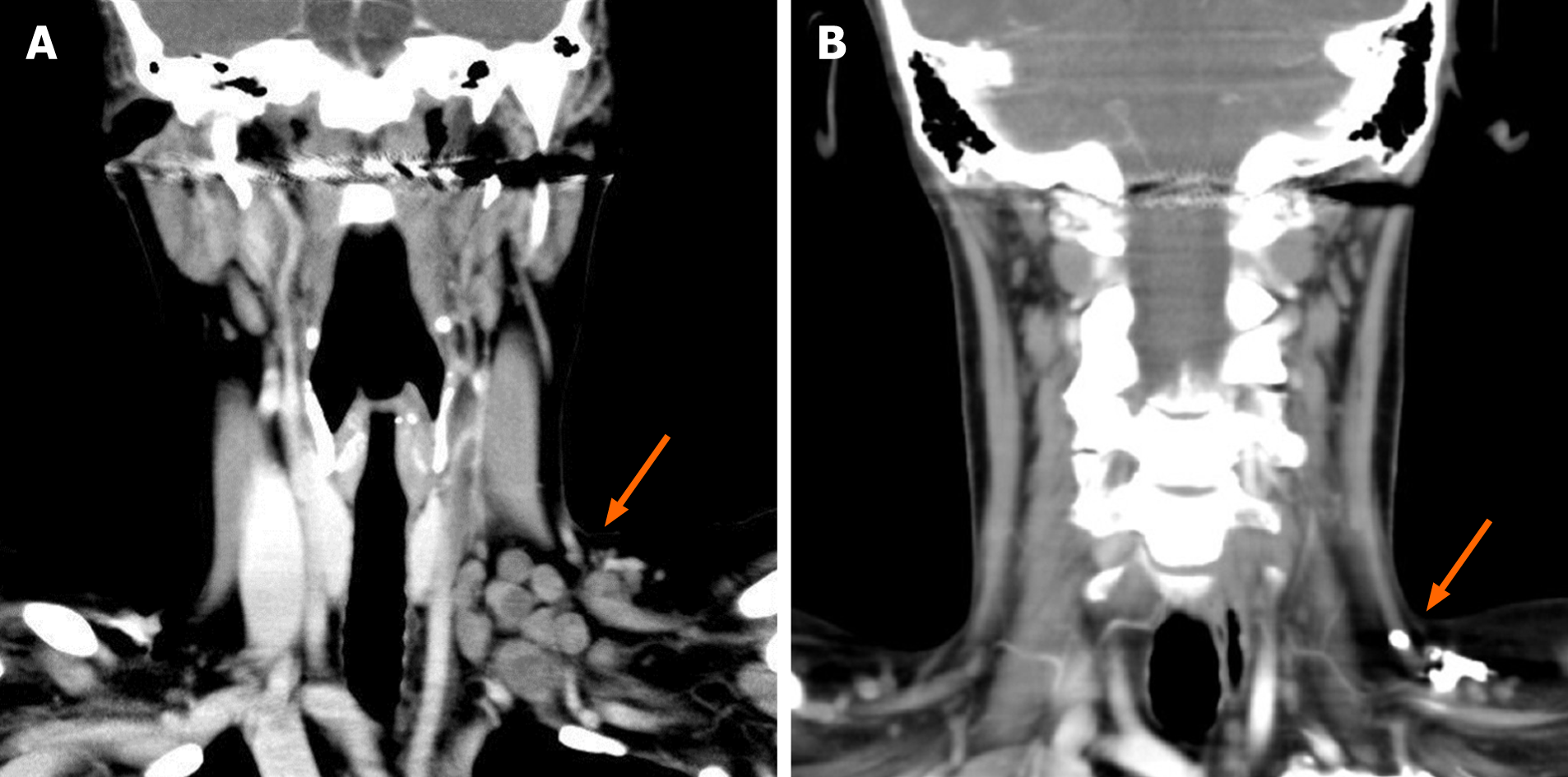
Figure 1 Computed tomography of the neck after intravenous injection of an iodine contrast agent.
A: Arrow pointing to swollen supraclavicular and cervical lymph nodes on the left; B: Complete remission of the initially swollen supraclavicular and cervical lymph nodes on the left.
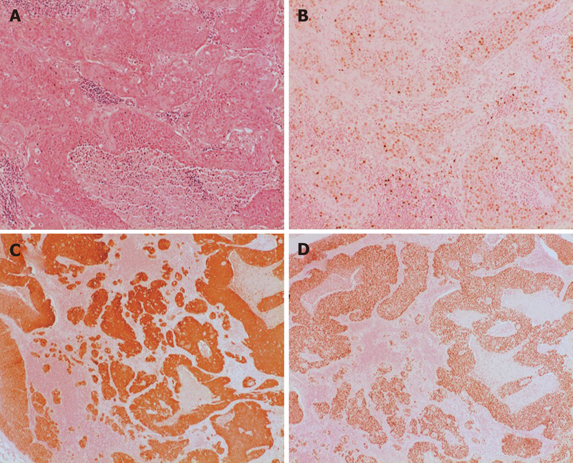
Figure 2 Lymph node with metastatic squamous cell carcinoma.
A: Hematoxylin-eosin staining; B: Ki67 staining; C: p16 staining; D: p40 staining.
Figure 3 Metastatic lymph nodes para-aortic region on the left.
A: Abdomen computed tomography after intravenous injection of an iodine contrast agent, arrow pointing on swollen lymph nodes; B: Fluorodeoxyglucose positron emission tomography signal positive lymph nodes in the para-aortic region.
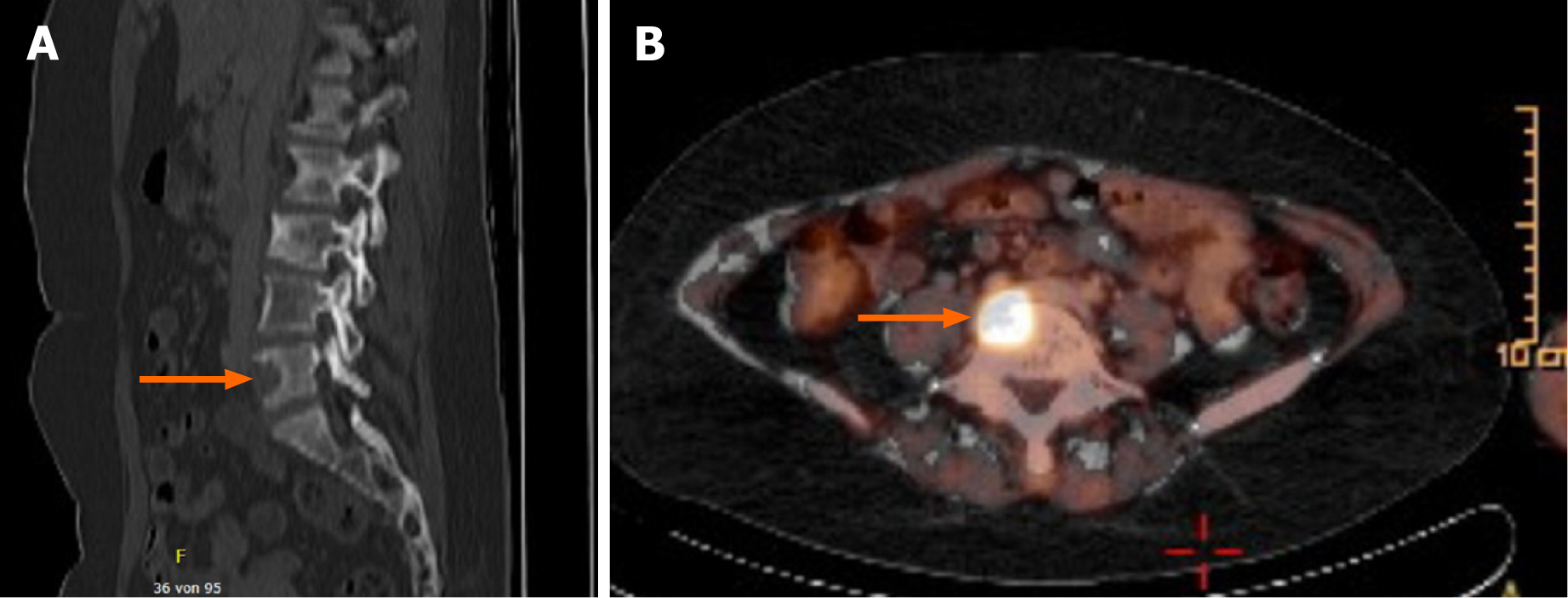
Figure 4 Bone metastasis in the fifth lumbar bone.
A: Computed tomography of the lumbar spine: arrow pointing to osseous destruction; B: Fluorodeoxyglucose positron emission tomography: Arrow pointing on signal positive bone metastasis.
- Citation: Große-Thie C, Maletzki C, Junghanss C, Schmidt K. Long-term survivor of metastatic squamous-cell head and neck carcinoma with occult primary after cetuximab-based chemotherapy: A case report. World J Clin Cases 2021; 9(24): 7092-7098
- URL: https://www.wjgnet.com/2307-8960/full/v9/i24/7092.htm
- DOI: https://dx.doi.org/10.12998/wjcc.v9.i24.7092


