Copyright
©The Author(s) 2021.
World J Clin Cases. Jan 16, 2021; 9(2): 509-515
Published online Jan 16, 2021. doi: 10.12998/wjcc.v9.i2.509
Published online Jan 16, 2021. doi: 10.12998/wjcc.v9.i2.509
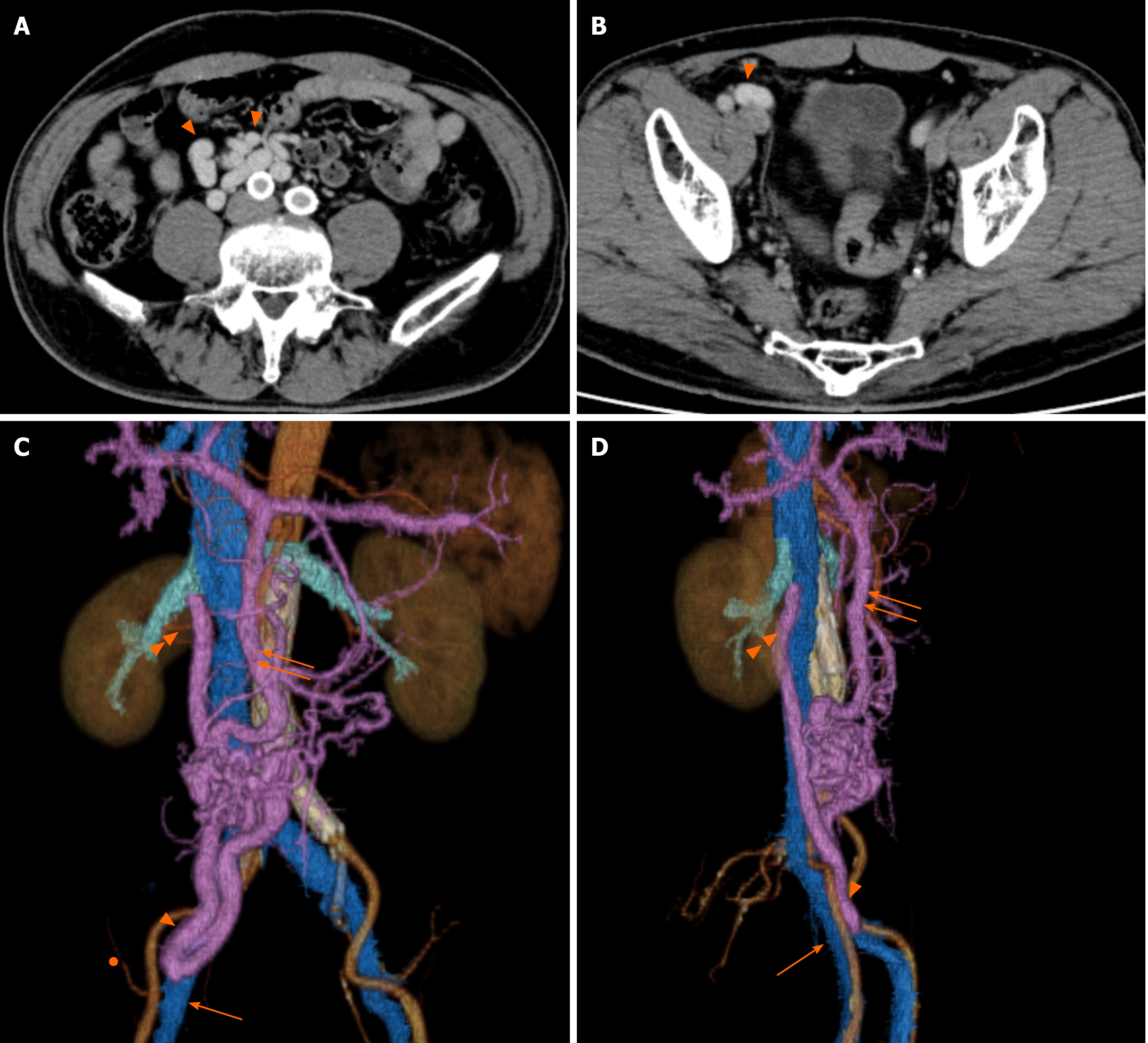
Figure 1 Computed tomography findings 7 years before surgery.
A: A portosystemic venous shunt vessel was evident (triangles); B: Portosystemic venous shunt vessel near the abdominal wall (triangle); C and D: A portosystemic venous shunt had formed between the ileocolic and testicular veins. Triangle: Shunt vessel; Double triangles: Testicular vein flowing into the inferior vena cava; Arrow: Femoral vein; Double arrows: Superior mesenteric vein; Circle: Inferior epigastric artery.
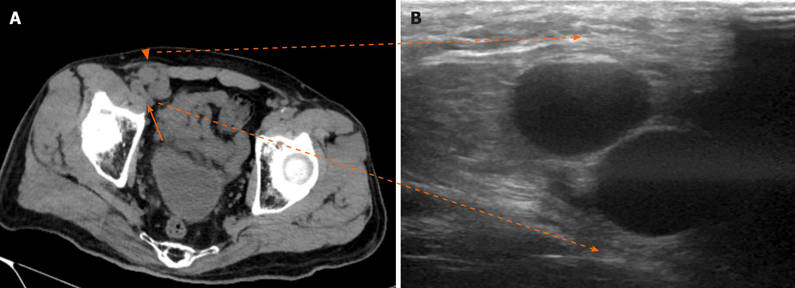
Figure 2 Preoperative computed tomography and ultrasound findings.
A: Preoperative computed tomography revealed a portosystemic venous shunt vessel located in the ventral part of the femoral vein (arrow) and entered the internal inguinal ring (triangle); B: Abdominal ultrasound detected the shunt vessel just under the groin.
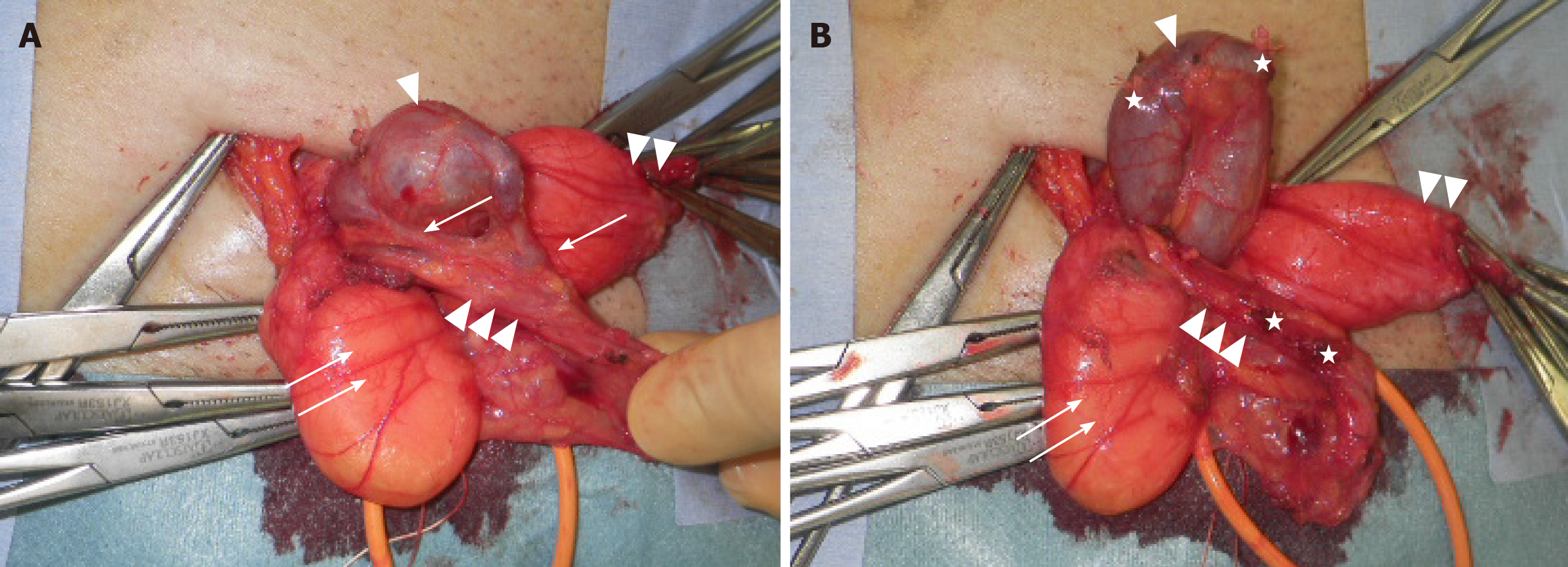
Figure 3 Photograph taken during surgery.
A: The expanded (2-cm-diameter) shunt vessel existing in the extraperitoneal cavity entered the inguinal canal through the internal inguinal ring. An indirect hernia sac and two short branches connecting the shunt vessel and the testicular vein were identified; B: Shunt vessel after cutting short branches. Triangle: Shunt vessel; Double triangles: Indirect hernia sac; Triple triangles: Vas deferens and testicular artery and vein; Arrow: Two short branches between the shunt vessel and testicular vein; Double arrows: Extraperitoneal fat; Stars: Cut-off stumps of ligated short branches.
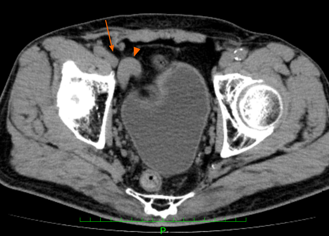
Figure 4 Postoperative computed tomography findings.
The computed tomography scans indicated that the shunt vessel was no longer located near the right internal inguinal ring, and it had separated from the femoral vein. Triangle: Shunt vessel; Arrow: Femoral vein.
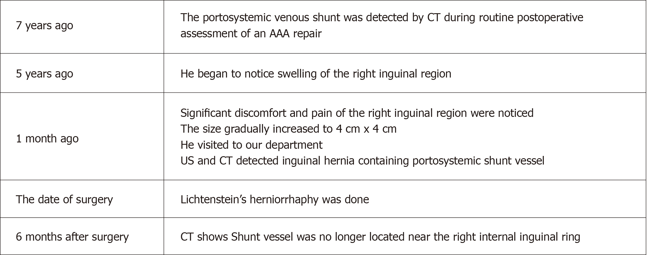
Figure 5 Timeline from diagnosis to treatment.
AAA: Abdominal aortic aneurysm; CT: Computed tomography; US: Ultrasound.
- Citation: Yura M, Yo K, Hara A, Hayashi K, Tajima Y, Kaneko Y, Fujisaki H, Hirata A, Takano K, Hongo K, Yoneyama K, Nakagawa M. Indirect inguinal hernia containing portosystemic shunt vessel: A case report. World J Clin Cases 2021; 9(2): 509-515
- URL: https://www.wjgnet.com/2307-8960/full/v9/i2/509.htm
- DOI: https://dx.doi.org/10.12998/wjcc.v9.i2.509


