Copyright
©The Author(s) 2021.
World J Clin Cases. Jan 16, 2021; 9(2): 482-488
Published online Jan 16, 2021. doi: 10.12998/wjcc.v9.i2.482
Published online Jan 16, 2021. doi: 10.12998/wjcc.v9.i2.482
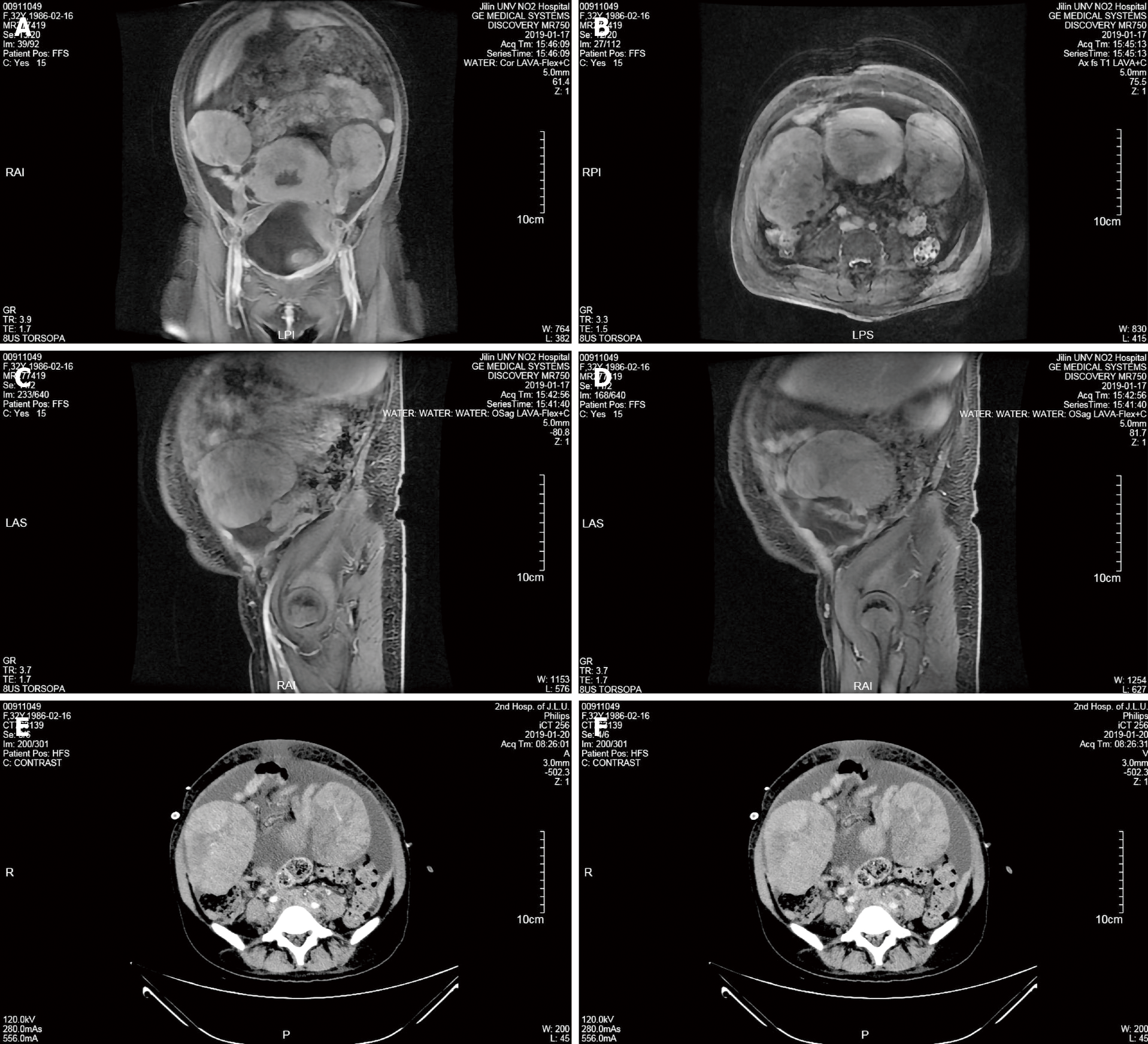
Figure 1 Magnetic resonance imaging and computed tomography imaging.
A and B: Coronal (A) and axial (B) images showed bilateral adnexal malignant tumors; C and D: Sagittal images showed masses in the left (C) and right (D) adnexal area; E and F: Computed tomography images of the arterial phase (E) and venous phase (F) showed bilateral adnexal malignant tumors.
Figure 2 Yellowish chyliform fluid drained from the abdomen.
- Citation: Xie F, Zhang LH, Yue YQ, Gu LL, Wu F. Double-hit lymphoma (rearrangements of MYC, BCL-2) during pregnancy: A case report. World J Clin Cases 2021; 9(2): 482-488
- URL: https://www.wjgnet.com/2307-8960/full/v9/i2/482.htm
- DOI: https://dx.doi.org/10.12998/wjcc.v9.i2.482


