Copyright
©The Author(s) 2021.
World J Clin Cases. Jan 16, 2021; 9(2): 429-435
Published online Jan 16, 2021. doi: 10.12998/wjcc.v9.i2.429
Published online Jan 16, 2021. doi: 10.12998/wjcc.v9.i2.429
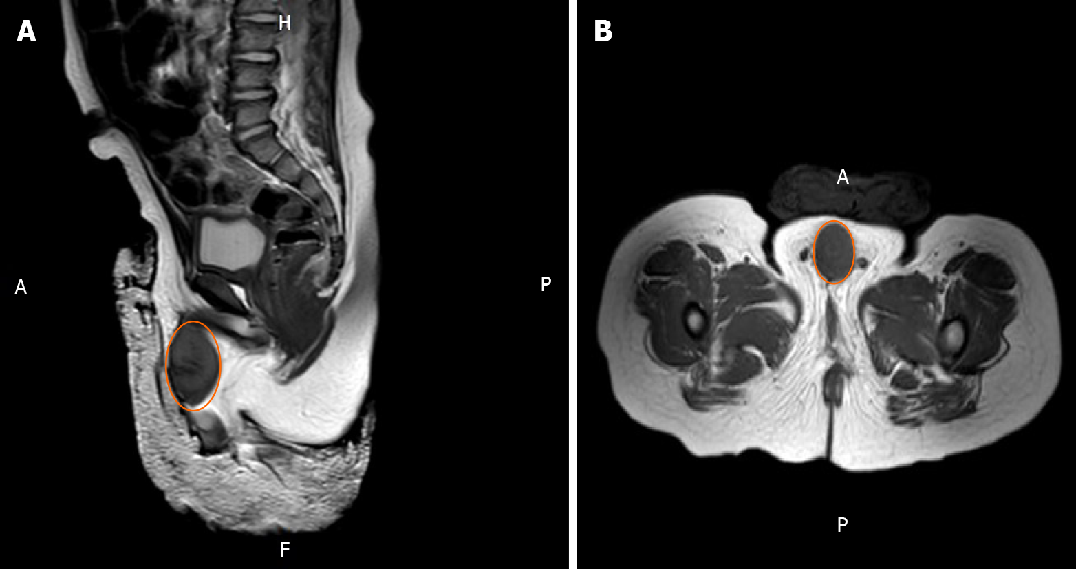
Figure 1 Magnetic resonance images of a tumor with rich blood supply encircling the cavernosum with a size of 3.
5 cm × 2.1 cm × 2.0 cm. A: Coronal image; B Axial image.
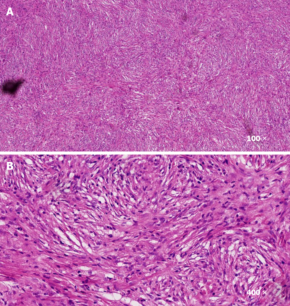
Figure 2 The tumor tissues at (A) 100 × magnification and (B) 400 × magnification stained with hematoxylin-eosin were rich in spindle cells with infiltration of inflammatory cells.
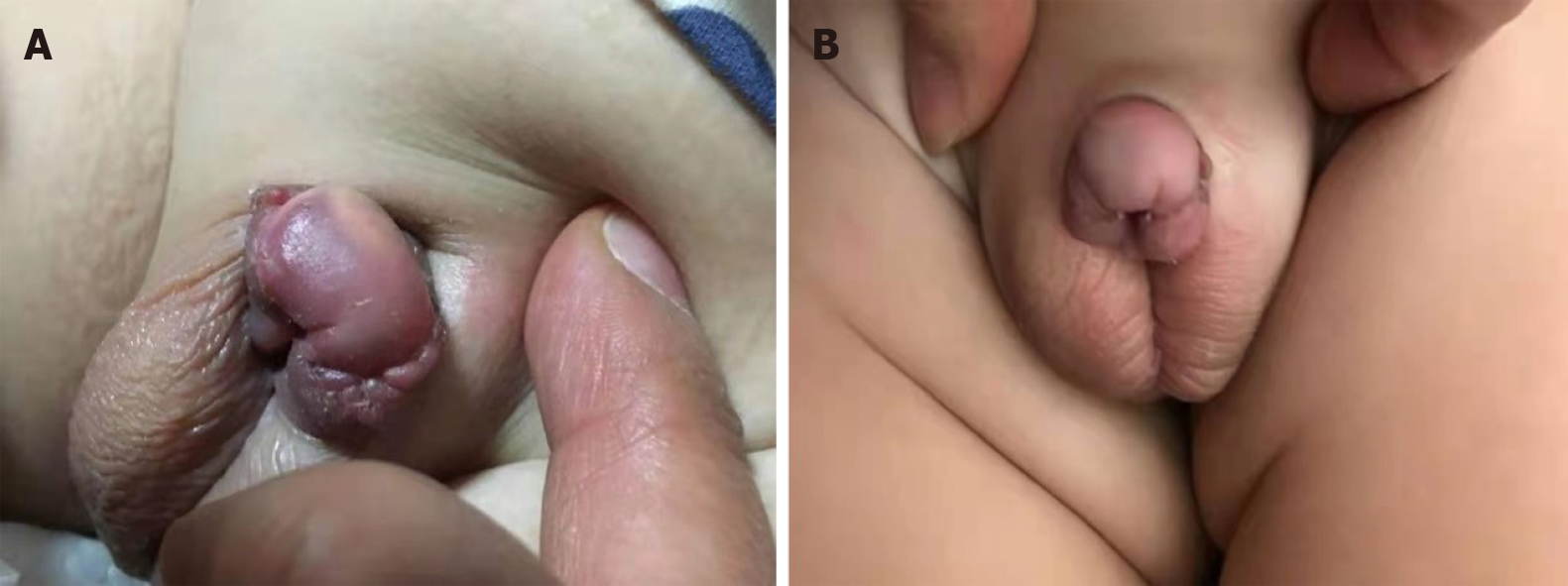
Figure 3 The tumor disappeared completely and the penis returned to normal (A and B).
- Citation: Liang Y, Gao HX, Tian RC, Wang J, Shan YH, Zhang L, Xie CJ, Li JJ, Xu M, Gu S. Inflammatory myofibroblastic tumor successfully treated with metformin: A case report and review of literature. World J Clin Cases 2021; 9(2): 429-435
- URL: https://www.wjgnet.com/2307-8960/full/v9/i2/429.htm
- DOI: https://dx.doi.org/10.12998/wjcc.v9.i2.429


