Copyright
©The Author(s) 2021.
World J Clin Cases. Jan 16, 2021; 9(2): 422-428
Published online Jan 16, 2021. doi: 10.12998/wjcc.v9.i2.422
Published online Jan 16, 2021. doi: 10.12998/wjcc.v9.i2.422
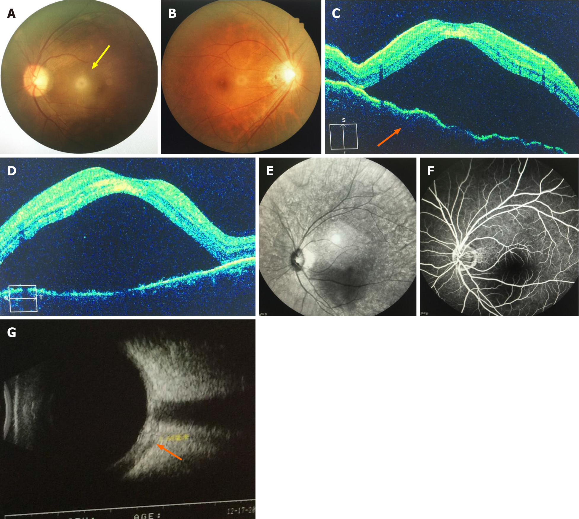
Figure 1 Imaging examinations.
A: Left eye fundus examination highlighting retinal edema and local retinal detachment; B: Right eye fundus examination without any remarkable findings; C: Left eye optical coherence tomography (OCT) showing choroidal folds and serous retinal detachment from the inferior to superior; D: Left eye OCT showing choroidal folds and serous retinal detachment from temporal to nasal; E: Red light free image of fluorescein angiography (FA) showing a dark area in the macula of the left eye; F: Final phases of FA showing no leakage in the choroid or retina of the left eye; G: Ultrasound showing “T-sign” (indicated by the orange arrow) in the left eye.
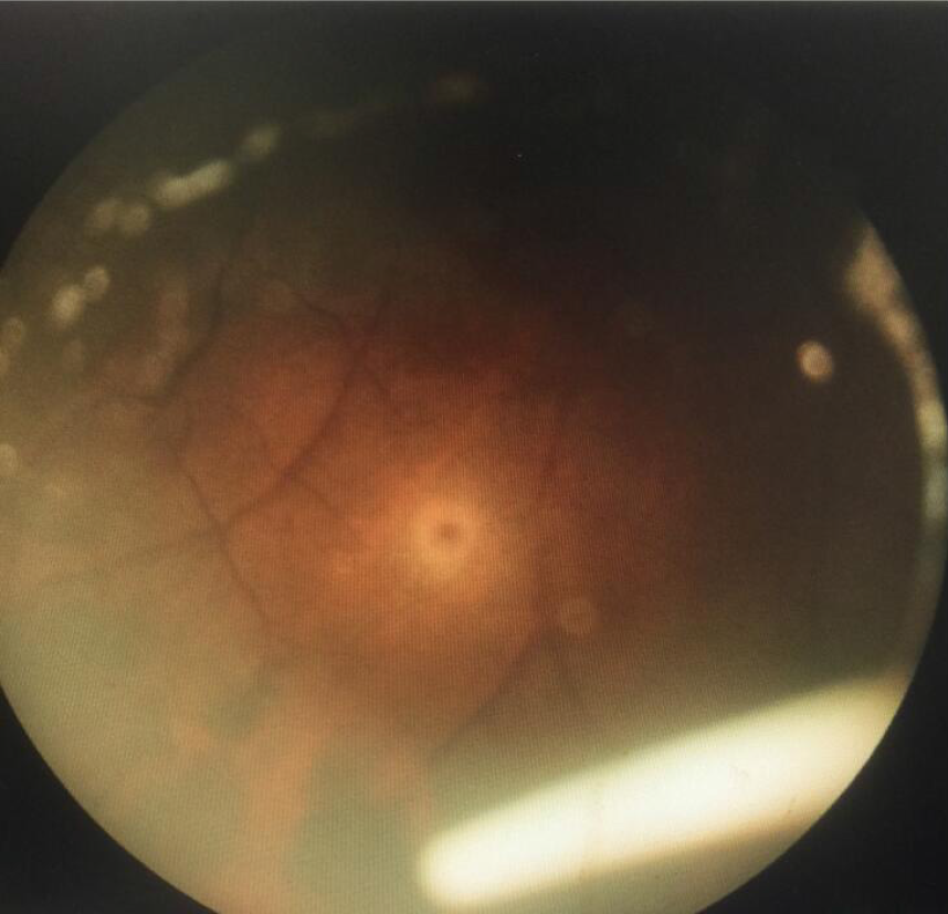
Figure 2 Ozurdex in the inferior of the vitreous cavity.
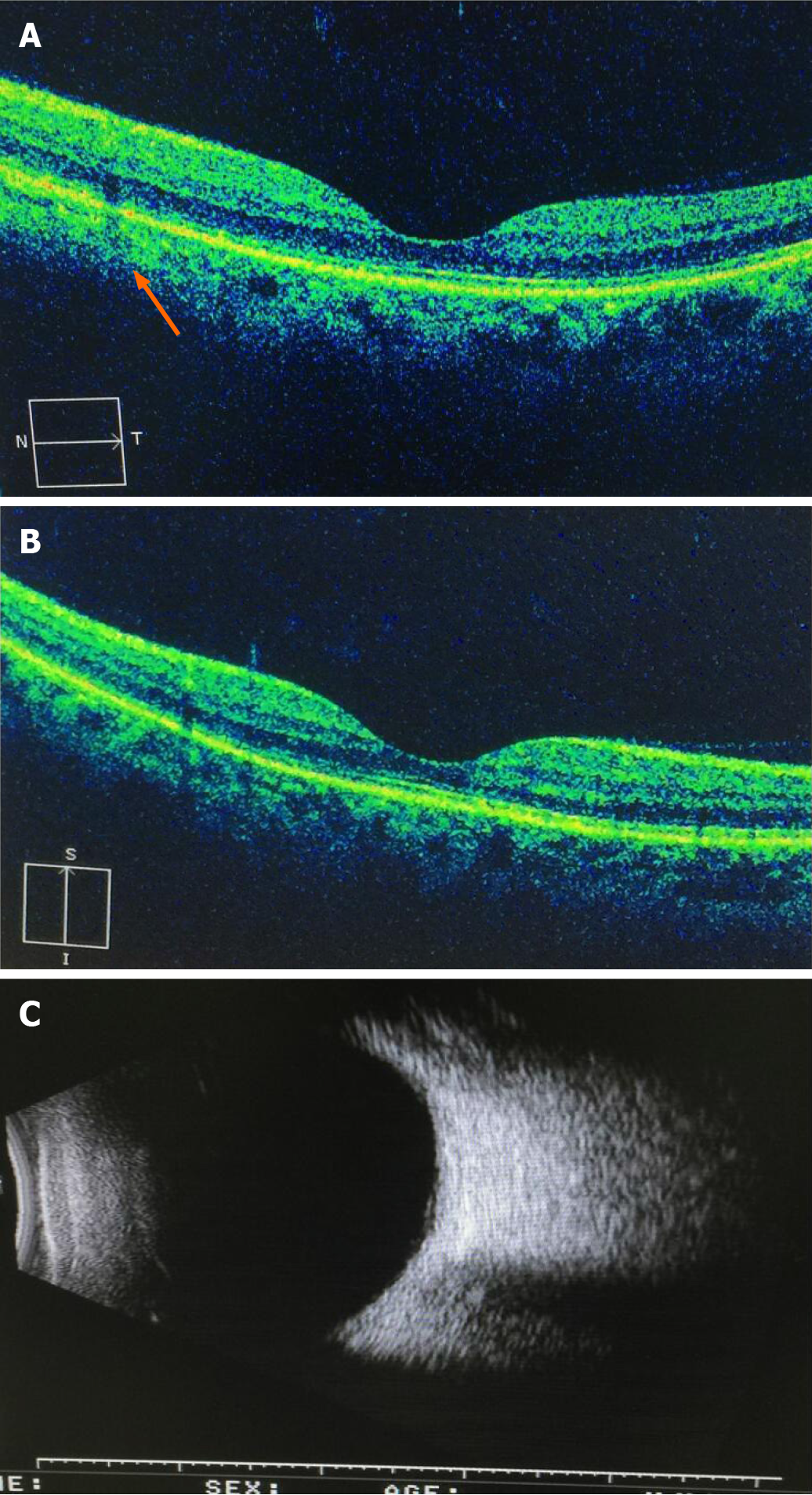
Figure 3 Optical coherence tomography and ultrasonography.
A: Retinal pigment epithelium and inner segment/outer segment layer in the nasal side of the macula were broken off (orange arrow); B: Total regression of the serous macular detachment of the left eye; C: Ultrasonography showed normal findings.
- Citation: Zhao YJ, Zou YL, Lu Y, Tu MJ, You ZP. Intravitreal dexamethasone implant — a new treatment for idiopathic posterior scleritis: A case report. World J Clin Cases 2021; 9(2): 422-428
- URL: https://www.wjgnet.com/2307-8960/full/v9/i2/422.htm
- DOI: https://dx.doi.org/10.12998/wjcc.v9.i2.422


