Copyright
©The Author(s) 2021.
World J Clin Cases. Jan 16, 2021; 9(2): 410-415
Published online Jan 16, 2021. doi: 10.12998/wjcc.v9.i2.410
Published online Jan 16, 2021. doi: 10.12998/wjcc.v9.i2.410
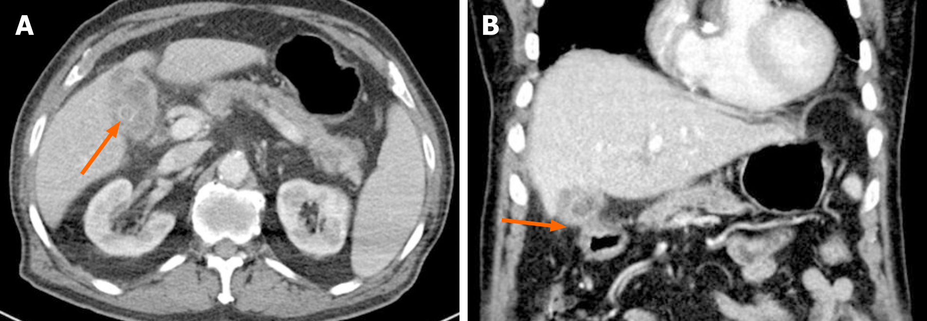
Figure 1 Radiologic images.
A: Computed tomography revealed a gallstone (arrow) and a non-dilated gallbladder with wall thickening, suggestive of chronic cholecystitis; B: The gallbladder was adherent to duodenum in coronal view.
Figure 2 Endoscopic images.
A: Esophagogastroduodenoscopy revealed an opening at the anterior wall of the duodenal bulb; B: Closer image of duodenal bulb; C: The opening was filled with a blood clot, suggestive of bleeding from cholecystoduodenal fistula.
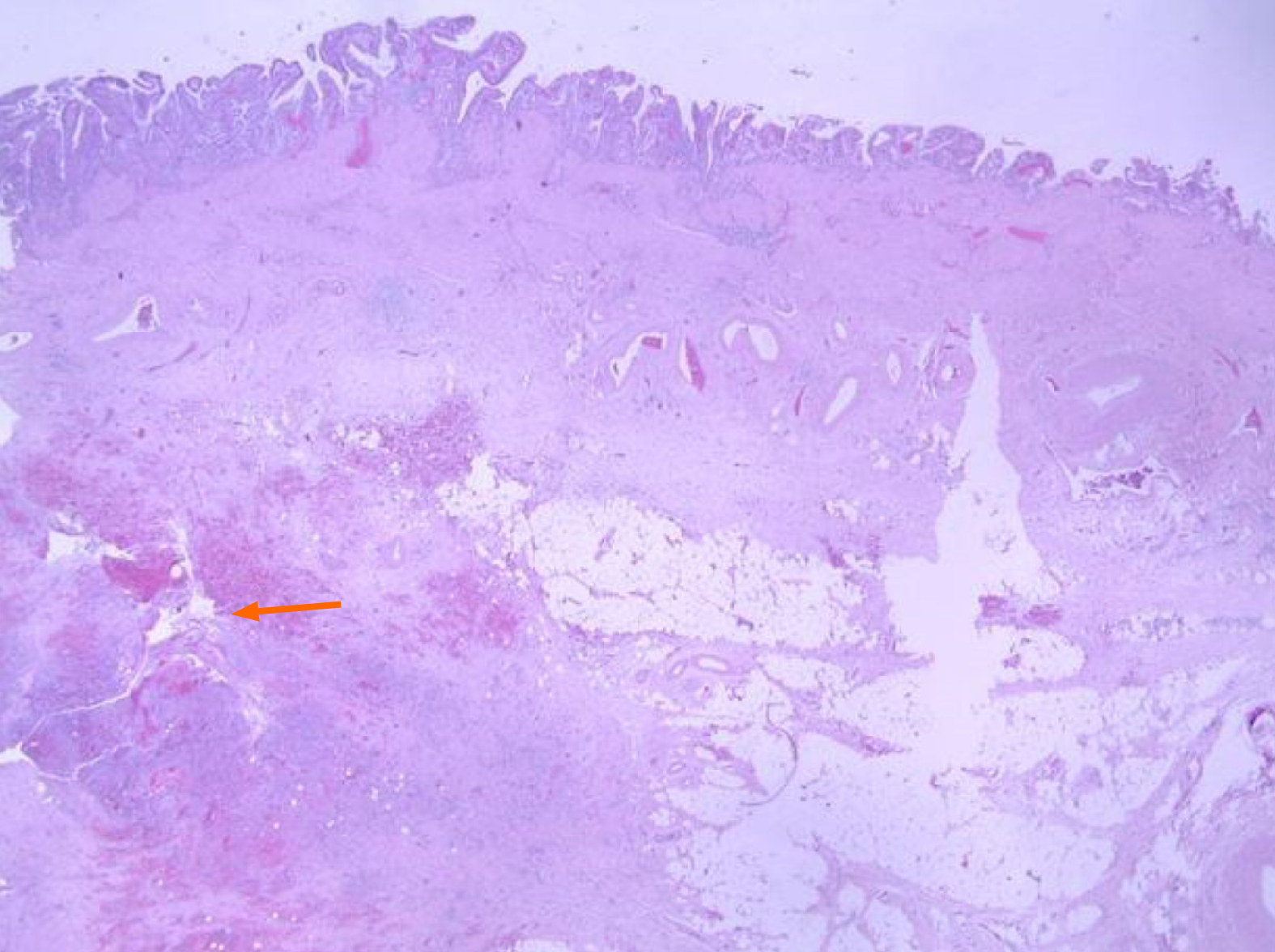
Figure 3 Pathologic image of resected gallbladder.
Microscopy shows the fistula in the lower portion of gallbladder mucosa (arrow, hematoxylin & eosin stain, × 15).
- Citation: Park JM, Kang CD, Kim JH, Lee SH, Nam SJ, Park SC, Lee SJ, Lee S. Cholecystoduodenal fistula presenting with upper gastrointestinal bleeding: A case report. World J Clin Cases 2021; 9(2): 410-415
- URL: https://www.wjgnet.com/2307-8960/full/v9/i2/410.htm
- DOI: https://dx.doi.org/10.12998/wjcc.v9.i2.410


