Copyright
©The Author(s) 2021.
World J Clin Cases. Jan 16, 2021; 9(2): 403-409
Published online Jan 16, 2021. doi: 10.12998/wjcc.v9.i2.403
Published online Jan 16, 2021. doi: 10.12998/wjcc.v9.i2.403
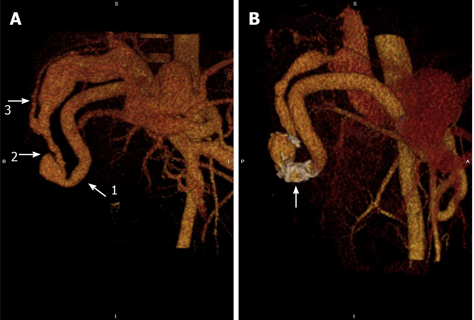
Figure 1 Contrast-enhanced computed tomography scan of the upper abdomen before and after coil embolization.
A: Three-dimensional computed tomography reconstruction demonstrated the hypertrophied right hepatic artery (11.8 mm, white arrow 1), the large arterioportal fistula (12 mm in diameter, white arrow 2), and the right portal vein (23.6 mm, white arrow 3). There is a round irregular vascular malformation (28 mm × 27.1 mm × 22.4 mm) around the fistula; B: There was a reduction in the right hepatic artery (9.5 mm), arterioportal fistula (5.8 mm) and right portal vein (11.1 mm) after embolization. The site of coil embolization in the distal part of the right hepatic artery is visible (white arrow).
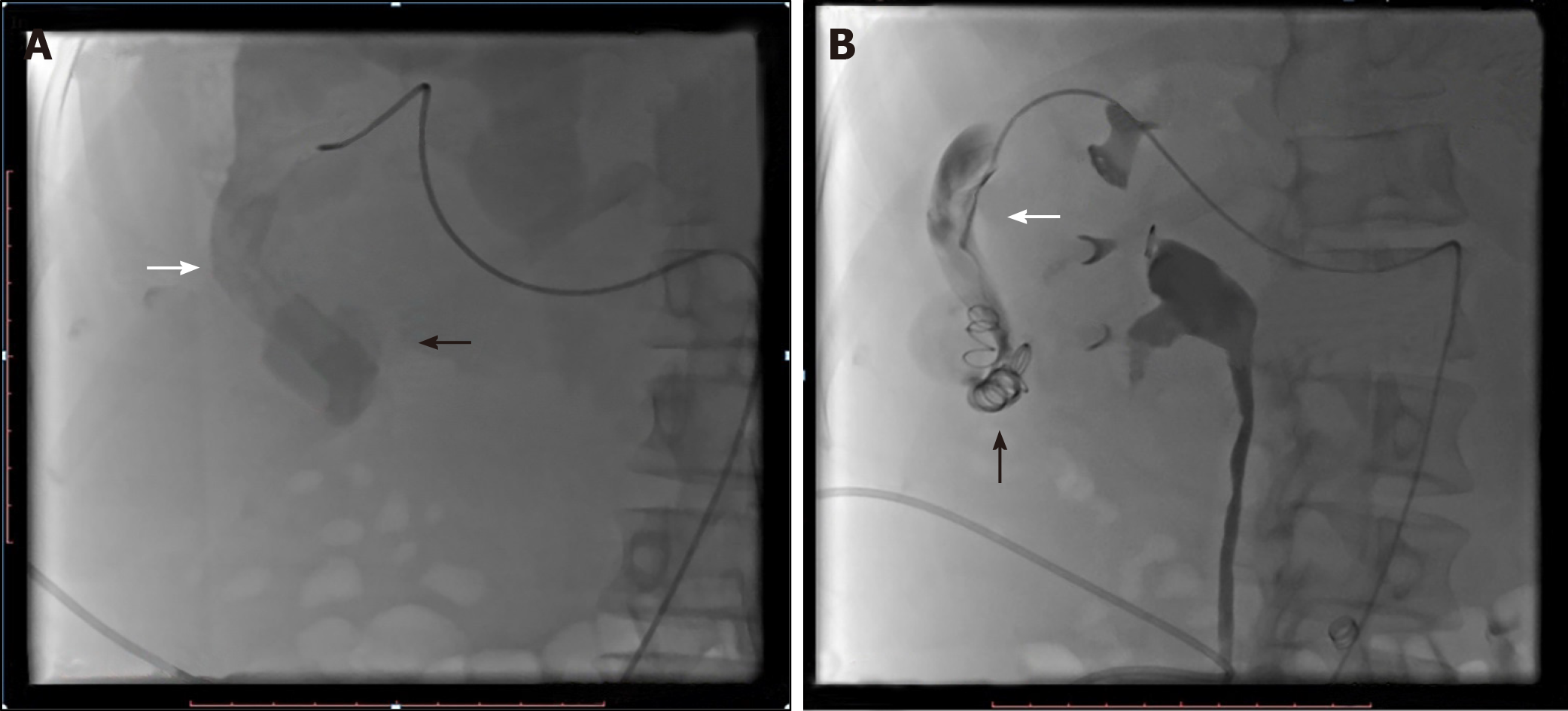
Figure 2 Selective right hepatic angiogram before and after coil embolization.
A: Angiography demonstrated the hypertrophied right hepatic artery (white arrow) and vascular malformation (black arrow) around the arterioportal fistula. B: Angiography showed the catheter in the enlarged right hepatic artery (white arrow). The distal segments of the right hepatic artery were occluded using two 12-3 and six 8-5 coils (black arrow).
- Citation: Stepanyan SA, Poghosyan T, Manukyan K, Hakobyan G, Hovhannisyan H, Safaryan H, Baghdasaryan E, Gemilyan M. Coil embolization of arterioportal fistula complicated by gastrointestinal bleeding after Caesarian section: A case report. World J Clin Cases 2021; 9(2): 403-409
- URL: https://www.wjgnet.com/2307-8960/full/v9/i2/403.htm
- DOI: https://dx.doi.org/10.12998/wjcc.v9.i2.403


