Copyright
©The Author(s) 2021.
World J Clin Cases. Jan 16, 2021; 9(2): 396-402
Published online Jan 16, 2021. doi: 10.12998/wjcc.v9.i2.396
Published online Jan 16, 2021. doi: 10.12998/wjcc.v9.i2.396
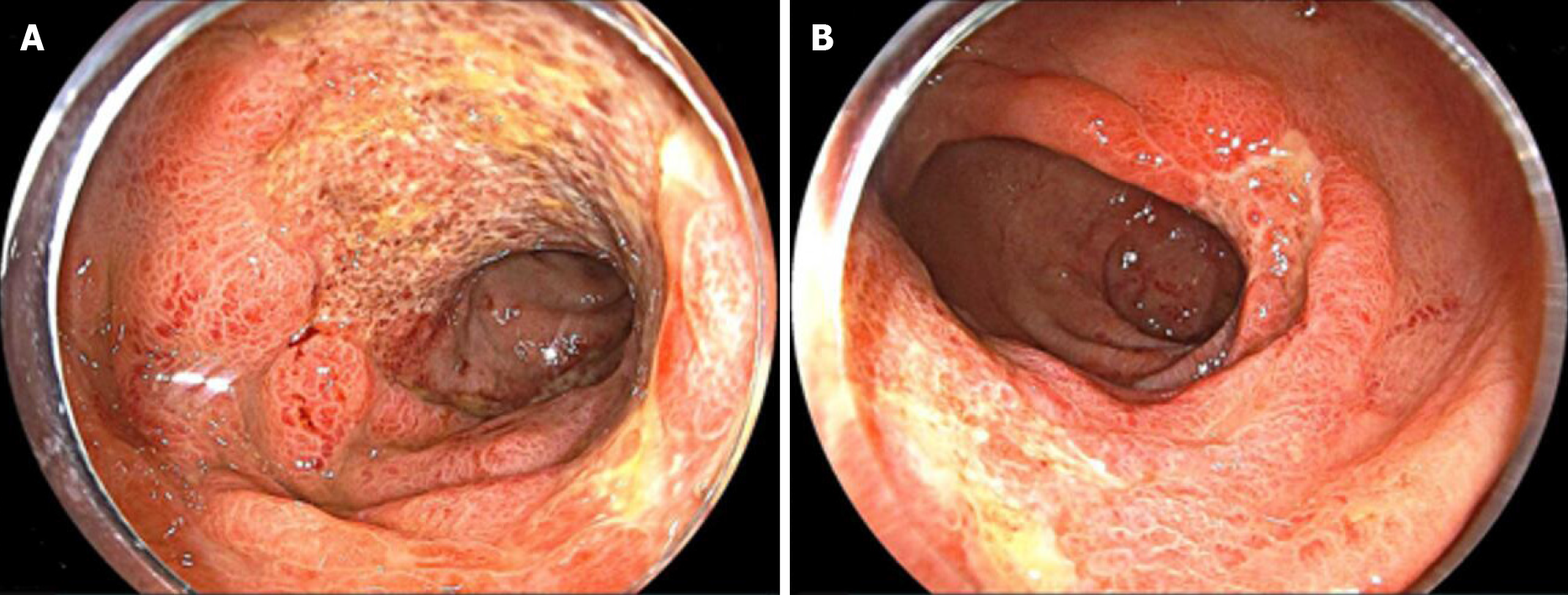
Figure 1 Colonoscopy findings at his first admission.
Colonoscopy showed diffuse ulceration, edema, and erythema from the descending colon to the rectum. These mucosal findings were most evident in the rectum above the peritoneal reflection. A: Colon; B: Rectum.
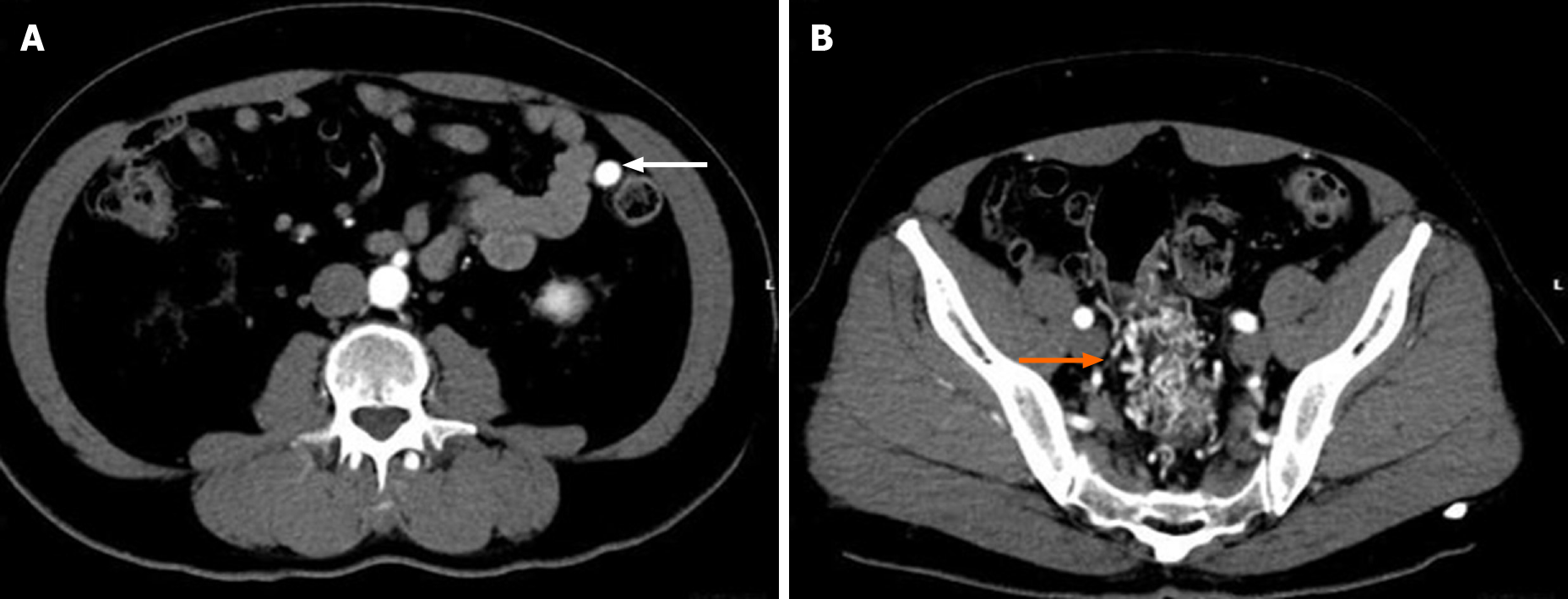
Figure 2 Images from contrast-enhanced abdominal computed tomography.
A contrast-enhanced abdominal computed tomography scan showed the nidus of small corkscrew and dilated vessels (orange arrow) and marked early inferior mesenteric vein enhancement (white arrow).
Figure 3 Image from computed tomography angiography.
Computed tomography angiography showed the same findings as contrast-enhanced abdominal computed tomography. The image showed the nidus of small corkscrew and dilated vessels arising from the superior rectal artery (orange arrowhead), which is a branch of the inferior mesenteric artery (orange arrow). Dilated inferior mesenteric veins were observed (white arrow).
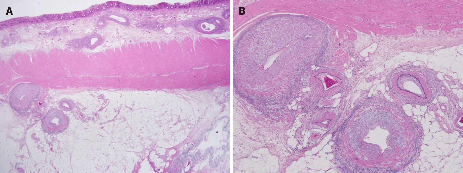
Figure 4 Histopathological findings of the surgical specimen.
Histopathological findings showed dilation of arteries and veins, which were close and mixed, consistent with an arteriovenous malformation. A: Victoria blue-hematoxylin-eosin × 12.5; B: Victoria blue-hematoxylin-eosin × 40.
Figure 5 Recent abdominal computed tomography.
A-D: Non-enhanced abdominal computed tomography images were taken before admission (A) and 2 years (B), 5 years (C), and 8 years (D) ago. The vessels around the rectosigmoid colon were not evident 8 years ago (D), but dilation of blood vessels around the rectosigmoid colon could have been detected 5 years ago (B and C).
- Citation: Kimura Y, Hara T, Nagao R, Nakanishi T, Kawaguchi J, Tagami A, Ikeda T, Araki H, Tsurumi H. Natural history of inferior mesenteric arteriovenous malformation that led to ischemic colitis: A case report. World J Clin Cases 2021; 9(2): 396-402
- URL: https://www.wjgnet.com/2307-8960/full/v9/i2/396.htm
- DOI: https://dx.doi.org/10.12998/wjcc.v9.i2.396


