Copyright
©The Author(s) 2021.
World J Clin Cases. Jan 16, 2021; 9(2): 379-388
Published online Jan 16, 2021. doi: 10.12998/wjcc.v9.i2.379
Published online Jan 16, 2021. doi: 10.12998/wjcc.v9.i2.379
Figure 1 Representative axial computed tomography images of the seventh thoracic vertebra.
A-C: A destructive lesion involving the T7 vertebral body and right pedicle was observed.
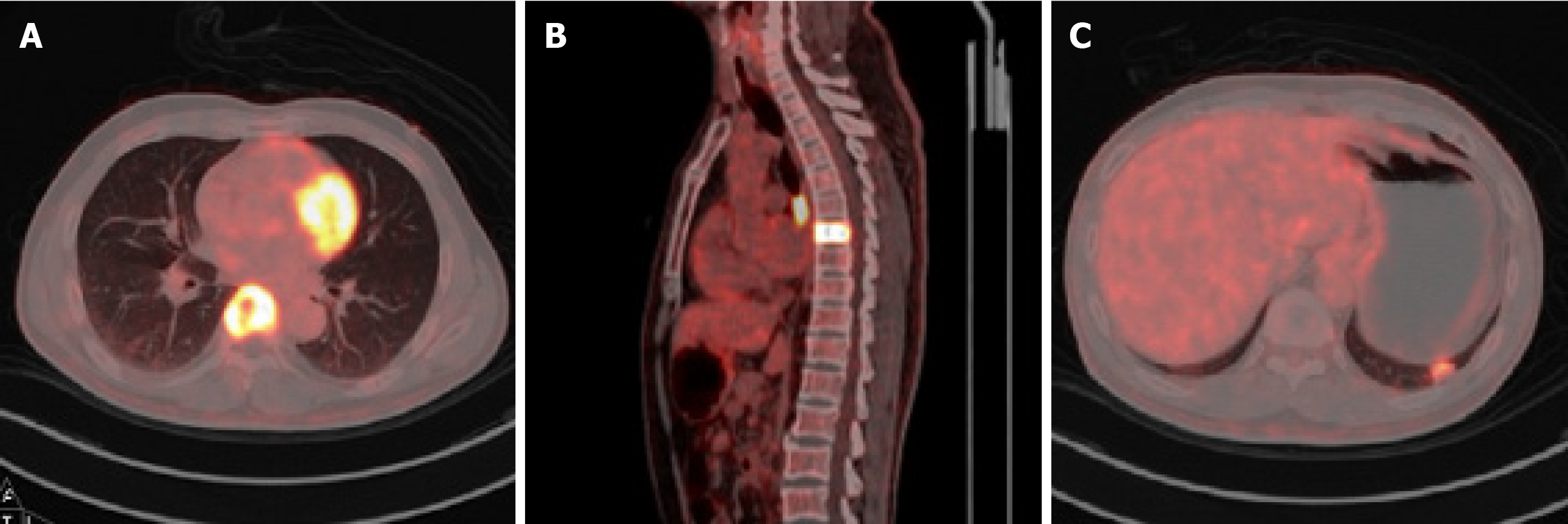
Figure 2 Representative positron emission tomography-computed tomography images of the patient.
A and B: Positron emission tomography-computed tomography (PET-CT) imaging of the seventh thoracic lesion. Its maximal standard uptake value (SUV) was 16.74; C: PET-CT scan of the left pulmonary mass (maximal SUV, 4.95).
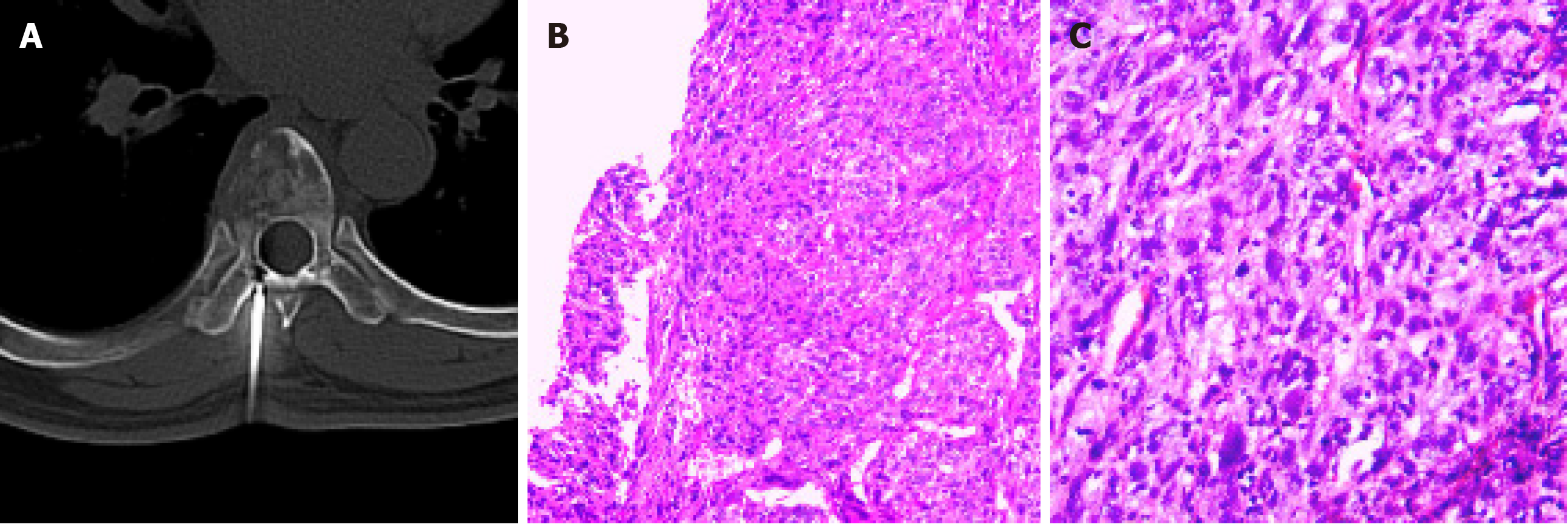
Figure 3 Pathological examinations of the patient.
A and B: Representative image of computed tomography-guided needle biopsy and the corresponding hematoxylin-eosin (H&E) staining result (magnification × 100); C: Postoperative H&E staining of the surgically removed tumor (magnification × 200).
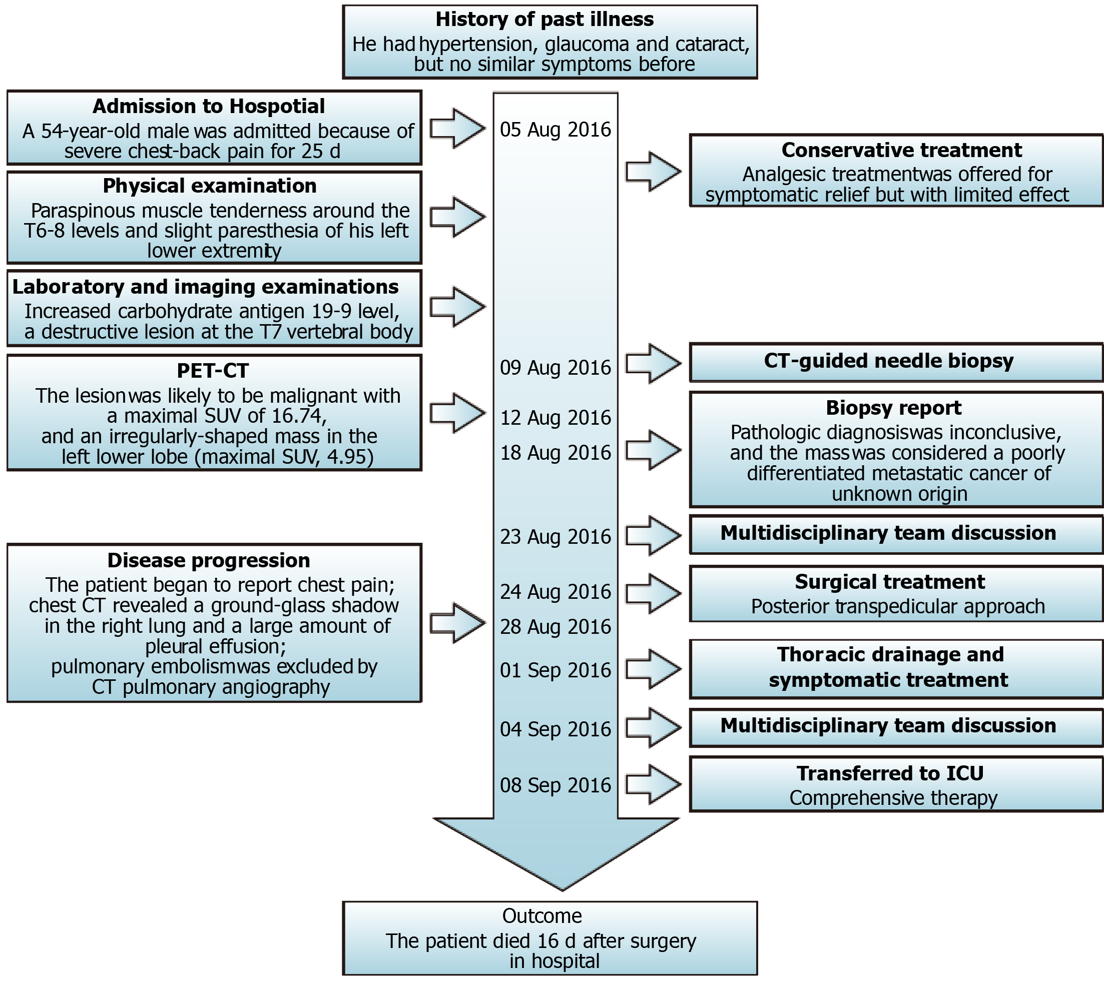
Figure 4 Information from this case report organized into a timeline.
PET-CT: Positron emission tomography-computed tomography; SUV: Standard uptake value; CT: Computed tomography; ICU: Intensive care unit.
- Citation: Li XM, Jin LB. Perioperative mortality of metastatic spinal disease with unknown primary: A case report and review of literature. World J Clin Cases 2021; 9(2): 379-388
- URL: https://www.wjgnet.com/2307-8960/full/v9/i2/379.htm
- DOI: https://dx.doi.org/10.12998/wjcc.v9.i2.379


