Copyright
©The Author(s) 2021.
World J Clin Cases. Jan 16, 2021; 9(2): 372-378
Published online Jan 16, 2021. doi: 10.12998/wjcc.v9.i2.372
Published online Jan 16, 2021. doi: 10.12998/wjcc.v9.i2.372
Figure 1 EUS visualization of varices and guidance of the angiotherapy.
A: The suspected esophageal varices are visualized by EUS. B: The injection of sclerosing /adhesive agent into the varix is shown. The needle tip is indicated by the white arrow.
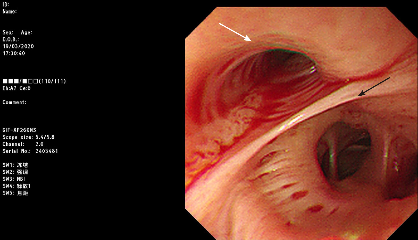
Figure 2 Ultra-thin gastroscopy used for suction of aspirated blood.
Ultra-thin gastroscope inserted into the endotracheal tube shows aspirated blood from both left (white arrow) and right bronchi, which are separated by the carina (black arrow).
- Citation: Wen TT, Liu ZL, Zeng M, Zhang Y, Cheng BL, Fang XM. Lateral position intubation followed by endoscopic ultrasound-guided angiotherapy in acute esophageal variceal rupture: A case report. World J Clin Cases 2021; 9(2): 372-378
- URL: https://www.wjgnet.com/2307-8960/full/v9/i2/372.htm
- DOI: https://dx.doi.org/10.12998/wjcc.v9.i2.372


