Copyright
©The Author(s) 2021.
World J Clin Cases. Jun 16, 2021; 9(17): 4433-4440
Published online Jun 16, 2021. doi: 10.12998/wjcc.v9.i17.4433
Published online Jun 16, 2021. doi: 10.12998/wjcc.v9.i17.4433
Figure 1 Retrospective review of previous lumbar spine magnetic resonance imaging findings revealed a cystic mass in the left extraforaminal space at the L5-S1 level.
A cystic mass along the path of left L5 spinal nerve.
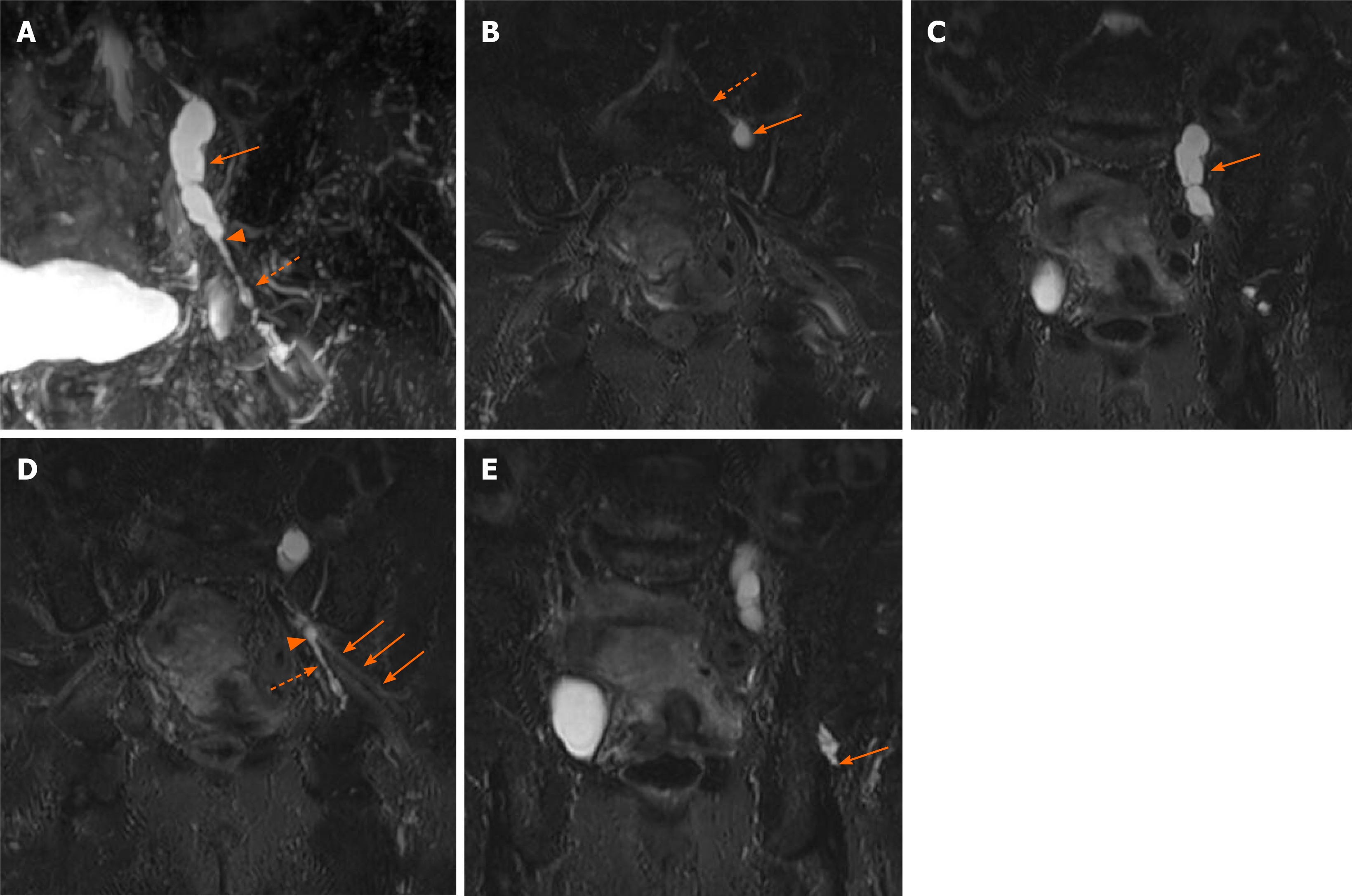
Figure 2 High-resolution magnetic resonance neurography.
A: Three-dimensional reconstructed maximum intensity projection images revealed an intraneural ganglion cyst in the articular branch of the hip joint. The cystic lesion had a tubular-like feature (dash arrow) from the termination of the articular branch to the level of the sciatic notch (arrow head) and a balloon-like feature (arrow) along the left L5 spinal nerve above the sciatic notch; B: The cyst (arrow) proximally extended to left L5 spinal nerve (dash arrow); C: The cyst lesion (arrow) had multifocal balloon-like dilatation; D: An intraneural ganglion cyst (dash arrow) in the articular branch of the hip joint combined with the left sciatic nerve (arrow) at the level of sciatic notch (arrow head); E: The cyst arose from the termination of the articular branch and seemed to connect with intra-articular space.
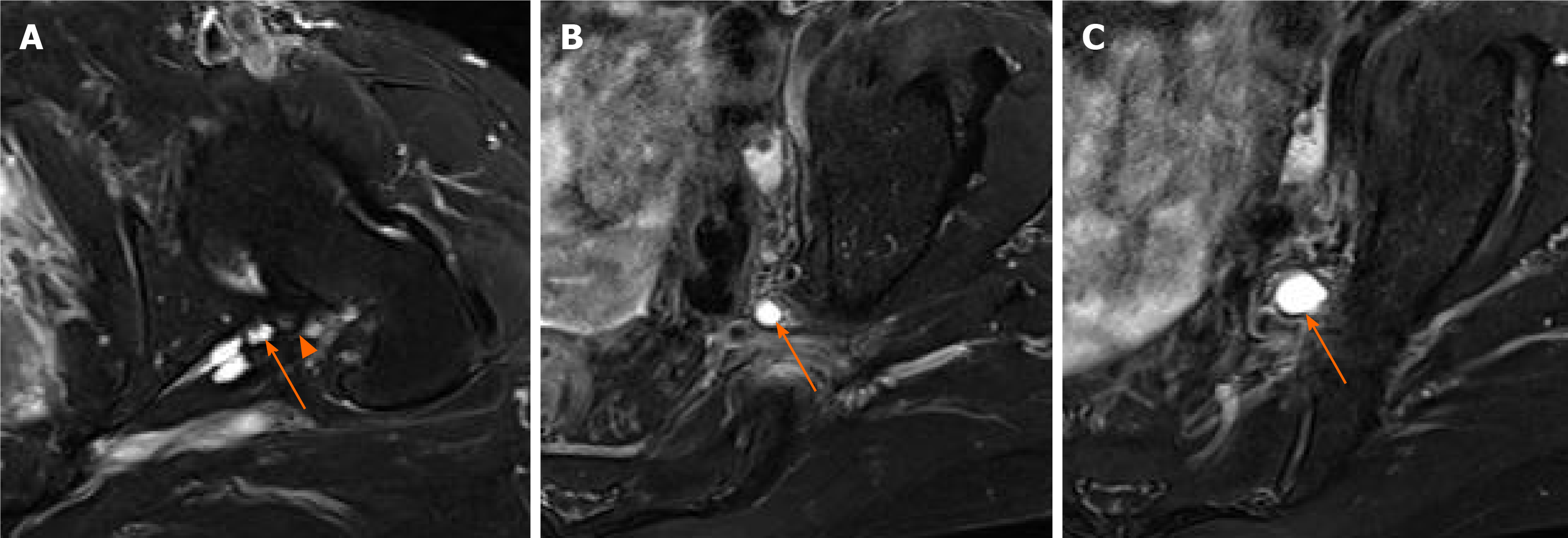
Figure 3 Magnetic resonance imaging.
A: A fat-suppressed T2-weighted axial image showing a degenerative change in the posterior labrum (arrowhead) and the paralabral cyst (arrow); B and C: Rostral to the level of the sciatic notch, the cyst (arrow) was more likely to be expansile and had a balloon-like feature.
- Citation: Lee JG, Peo H, Cho JH, Kim DH. Intraneural ganglion cyst of the lumbosacral plexus mimicking L5 radiculopathy: A case report. World J Clin Cases 2021; 9(17): 4433-4440
- URL: https://www.wjgnet.com/2307-8960/full/v9/i17/4433.htm
- DOI: https://dx.doi.org/10.12998/wjcc.v9.i17.4433


