Copyright
©The Author(s) 2021.
World J Clin Cases. Jun 16, 2021; 9(17): 4408-4414
Published online Jun 16, 2021. doi: 10.12998/wjcc.v9.i17.4408
Published online Jun 16, 2021. doi: 10.12998/wjcc.v9.i17.4408
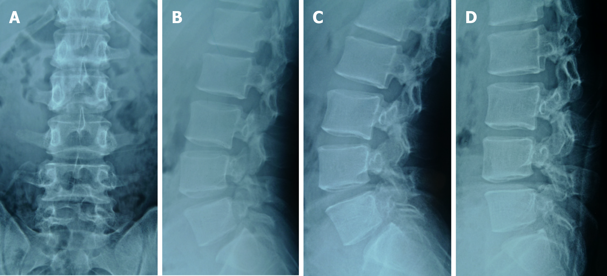
Figure 1 Lumbar radiographs.
A: Anteroposterior radiograph; B: Lateral radiograph showed L2-L5 spondylolysis; C: Lateral radiograph of lumbar extension position; D: Lateral radiograph of lumbar flexion position. Note that lumbar dynamic radiographs showed no instability.
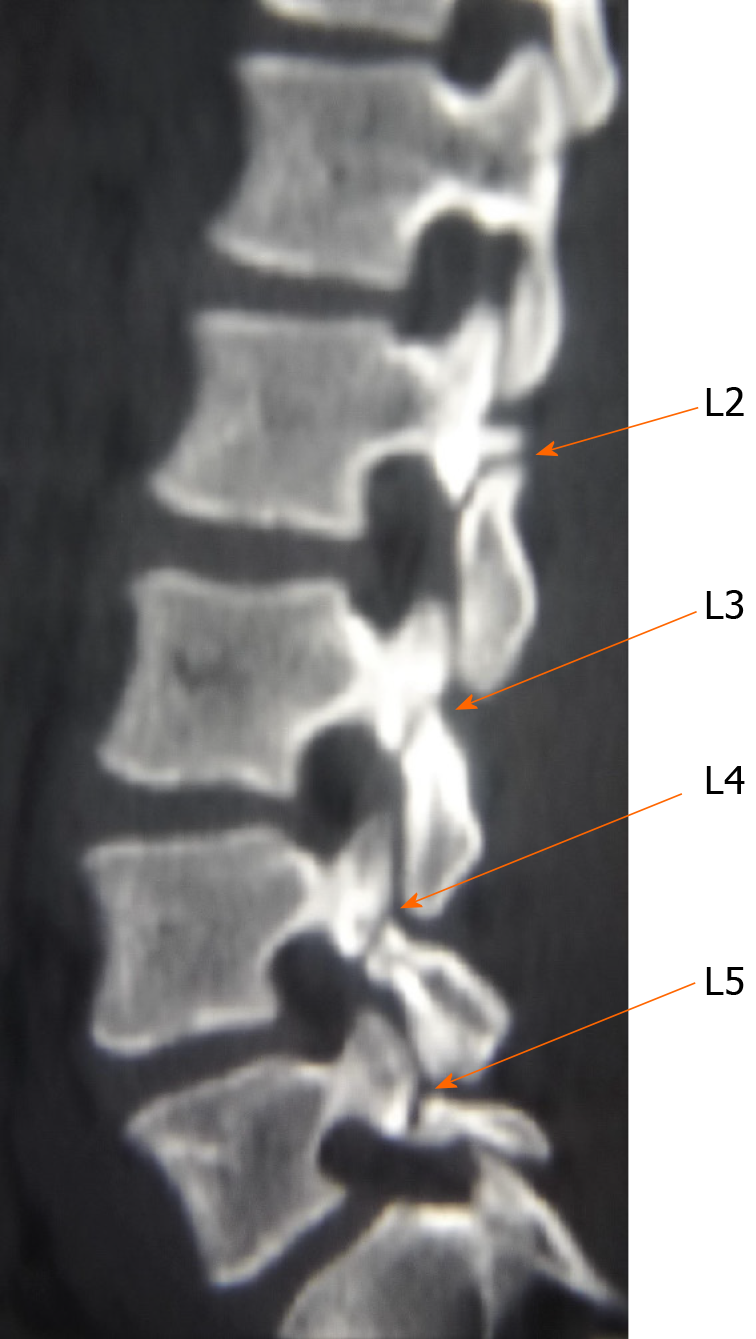
Figure 2 Two-dimensional computed tomography scan shows lumbar spondylolysis at bilateral L2-L5 levels.
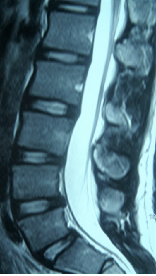
Figure 3 Magnetic resonance imaging showed no signs of lumbar disc degeneration.
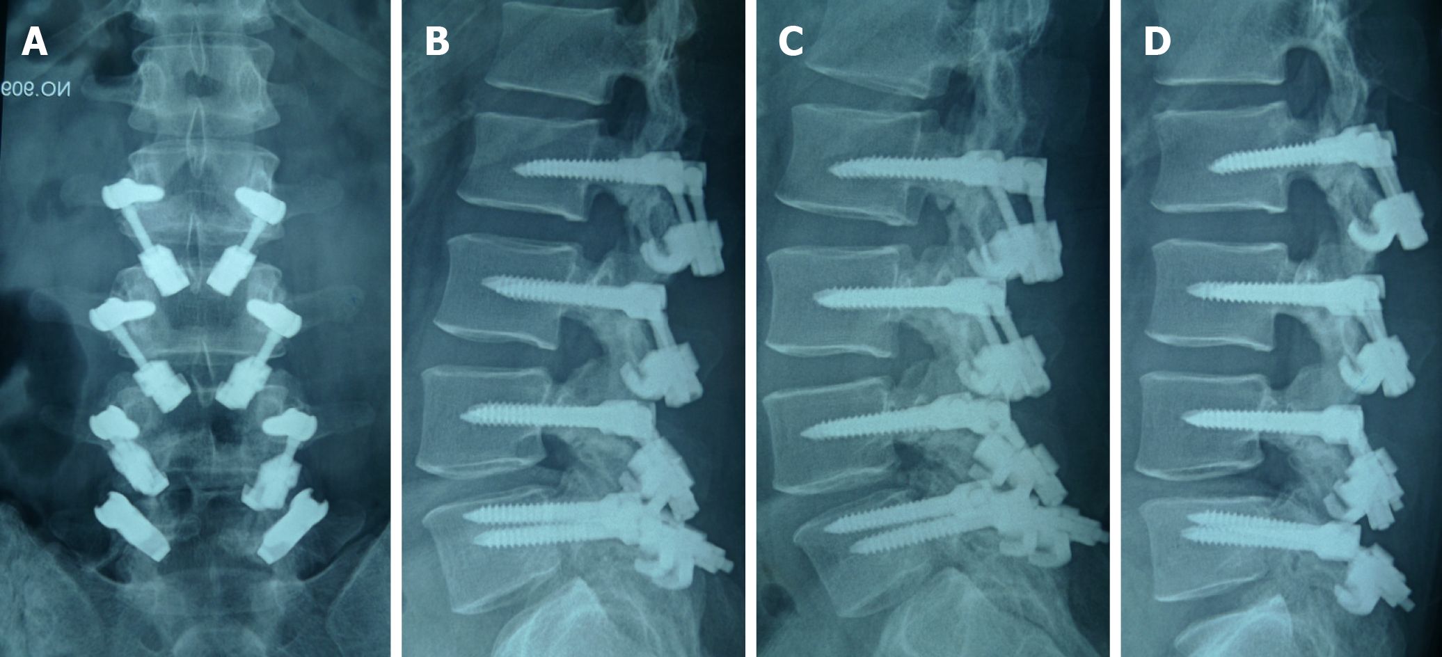
Figure 4 Lumbar dynamic radiographs after operation showed that lumbar movement was retained.
A: Anteroposterior radiograph; B: Lateral radiograph; C: Lateral radiograph of lumbar extension position; D: Lateral radiograph of lumbar flexion position. Note that lumbar dynamic radiographs show that movement was retained.
Figure 5 Computed tomography scan showed bone healing in all eight lytic defects at L2-L5.
A: L2; B: L3; C: L4; D: L5.
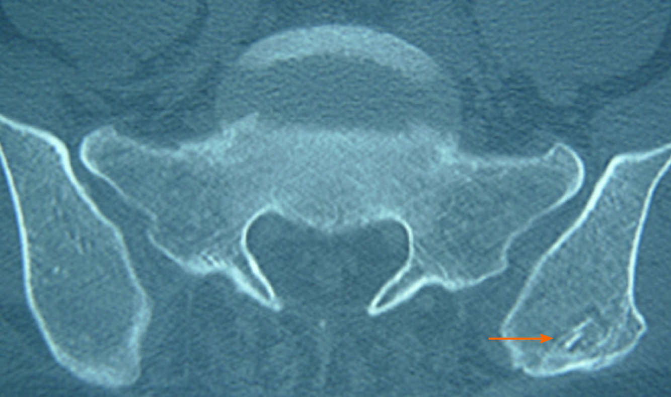
Figure 6 Donor site of iliac crest was filled with allogeneic bone, which resulted in osteogenesis (arrow).
- Citation: Li DM, Peng BG. Surgical treatment of four segment lumbar spondylolysis: A case report . World J Clin Cases 2021; 9(17): 4408-4414
- URL: https://www.wjgnet.com/2307-8960/full/v9/i17/4408.htm
- DOI: https://dx.doi.org/10.12998/wjcc.v9.i17.4408


