Copyright
©The Author(s) 2021.
World J Clin Cases. Jun 16, 2021; 9(17): 4400-4407
Published online Jun 16, 2021. doi: 10.12998/wjcc.v9.i17.4400
Published online Jun 16, 2021. doi: 10.12998/wjcc.v9.i17.4400
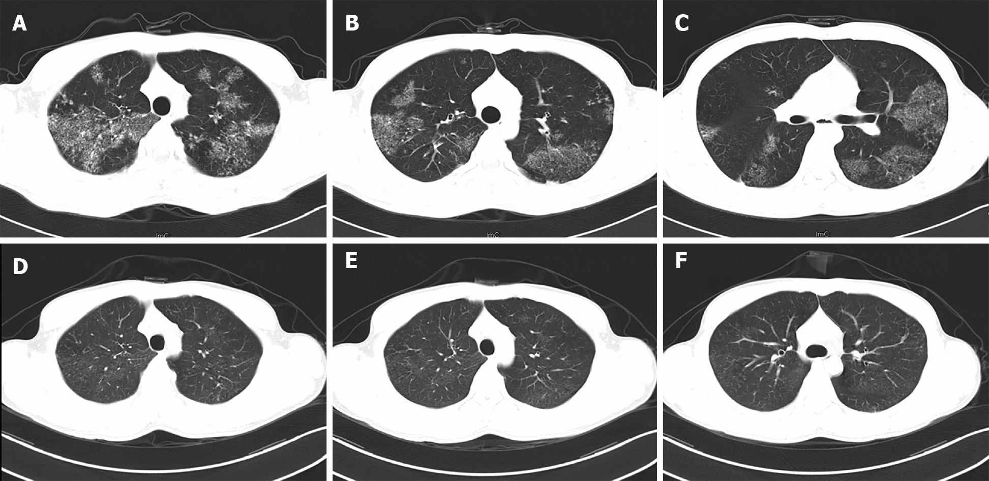
Figure 1 High-resolution computerized tomography scans of the chest.
A-C: Images before bronchoalveolar lavage and anti-tuberculosis treatment; D-F: Images after bronchoalveolar lavage and 6 mo of anti-tuberculosis treatment.
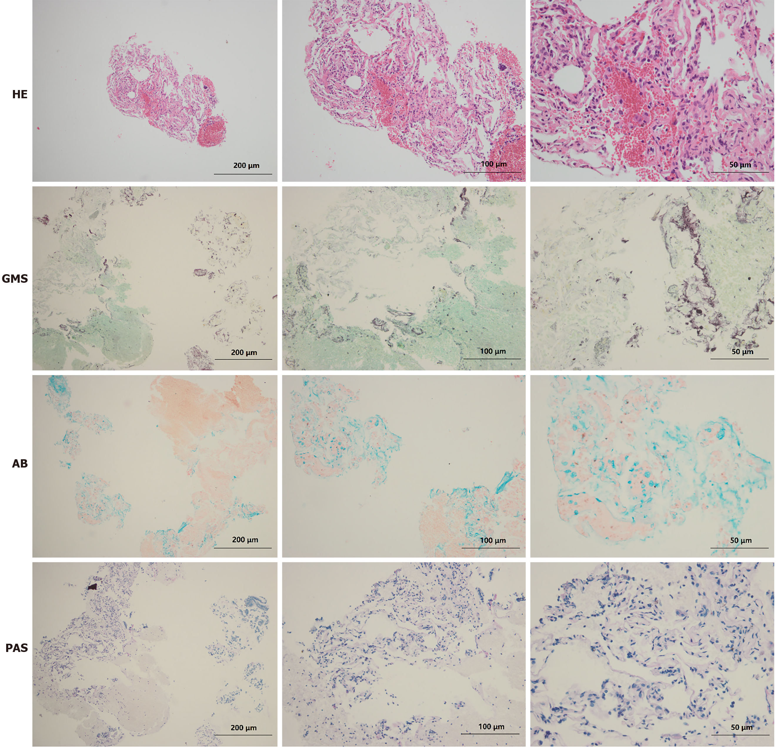
Figure 2 Histological examinations of lung biopsy.
HE: Hematoxylin-eosin staining; GMS: Gomori's methenamine silver staining; AB: Alcian blue staining; PAS: Periodic acid-Schiff staining.
- Citation: Bai H, Meng ZR, Ying BW, Chen XR. Pulmonary alveolar proteinosis complicated with tuberculosis: A case report. World J Clin Cases 2021; 9(17): 4400-4407
- URL: https://www.wjgnet.com/2307-8960/full/v9/i17/4400.htm
- DOI: https://dx.doi.org/10.12998/wjcc.v9.i17.4400


