Copyright
©The Author(s) 2021.
World J Clin Cases. Jun 16, 2021; 9(17): 4388-4394
Published online Jun 16, 2021. doi: 10.12998/wjcc.v9.i17.4388
Published online Jun 16, 2021. doi: 10.12998/wjcc.v9.i17.4388
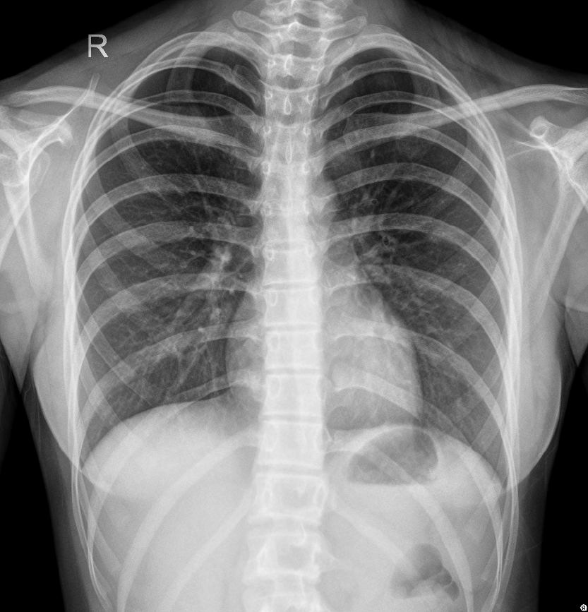
Figure 1 Chest X-ray showing thickness of texture in both lungs.
Figure 2 Chest computed tomography A: At the plane of the T2/T3 vertebrae; B: A coronal reconstruction showing a nodular mass in the lumen of the trachea.
Figure 3 Bronchoscopy image showing a mid-tracheal, pedicled dumbbell-shaped mass with small blood vessels on its surface.
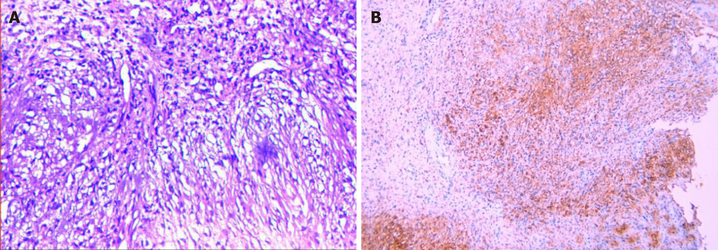
Figure 4 Pathological and immunohistochemical images.
A: Showing long, shuttle-shaped tumor cells with an uneven distribution and fenestrated arrangement in some areas (hematoxylin and eosin, magnification, × 100); B: Positive immunohistochemical staining of S-100 protein (magnification, × 100).
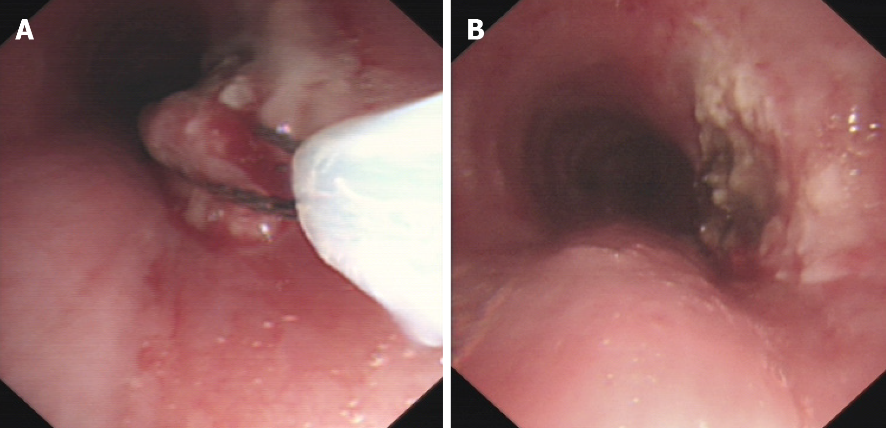
Figure 5 Bronchoscopy images.
A: Removal of the mass by high-frequency electrosurgery; B: Residual anatomy after mass removal.
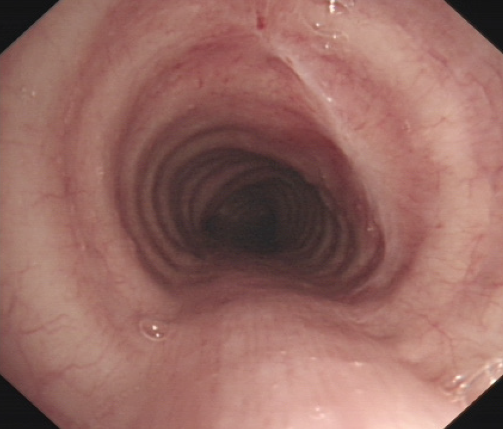
Figure 6 Bronchoscopy image showing slight scarring of the trachea at the site of the original lesion.
- Citation: Huang HR, Li PQ, Wan YX. Primary intratracheal schwannoma misdiagnosed as severe asthma in an adolescent: A case report. World J Clin Cases 2021; 9(17): 4388-4394
- URL: https://www.wjgnet.com/2307-8960/full/v9/i17/4388.htm
- DOI: https://dx.doi.org/10.12998/wjcc.v9.i17.4388


