Copyright
©The Author(s) 2021.
World J Clin Cases. Jun 16, 2021; 9(17): 4373-4380
Published online Jun 16, 2021. doi: 10.12998/wjcc.v9.i17.4373
Published online Jun 16, 2021. doi: 10.12998/wjcc.v9.i17.4373
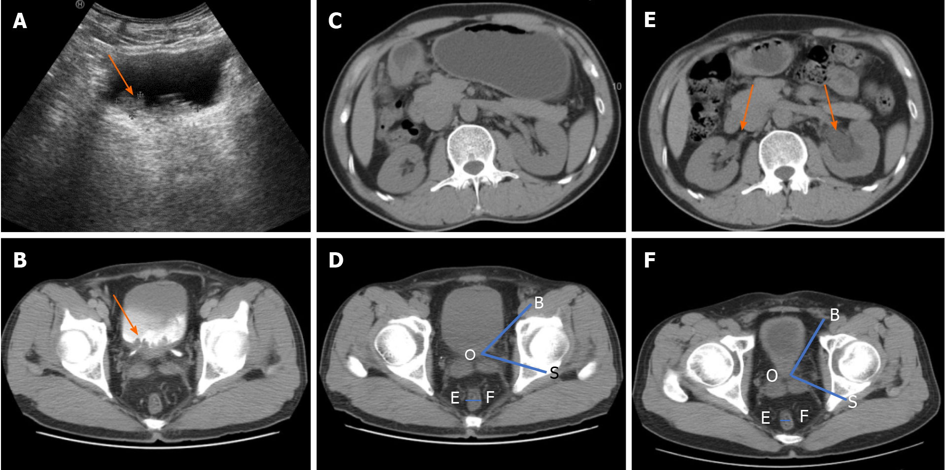
Figure 1 Imaging findings of cystitis glandularis with pelvic lipomatosis.
A: Ultrasound examination showed that the mass was located in the triangular region of the bladder, with a burr-like protrusion and irregular thickened wall of the triangular region of the bladder; B: On first admission, computed tomography (CT) of the urinary system suggested thickening of the bladder wall, mainly on the posterior wall and the triangle area, and the mastoid process was observed inside the cavity. Mild to moderate continuous enhancement was noted in the delayed phase; C: Urinary CT scan for the first time revealed no hydrops in the renal pelvis and ureter; D: Angle between the bladder and seminal vesicle (ABS) in the axial cross-section (∠BOS) and the axial cross-sectional diameter of the rectum (EF) in CT at the first visit. The bladder morphology was elliptical; E: CT was reviewed 3 mo after the second transurethral resection of bladder tumour (TUR-BT). The pelvises, calyces, and ureters on both sides dilated, especially on the left side; F: CT review 3 mo after the second TUR-BT: ABS in the axial cross-section (∠BOS) became larger and the axial cross-sectional diameter of the rectum (EF) became smaller. The adipose density around the bladder and around the rectum increased, the rectum was compressed, and the bladder shape was inversely pear-shaped.
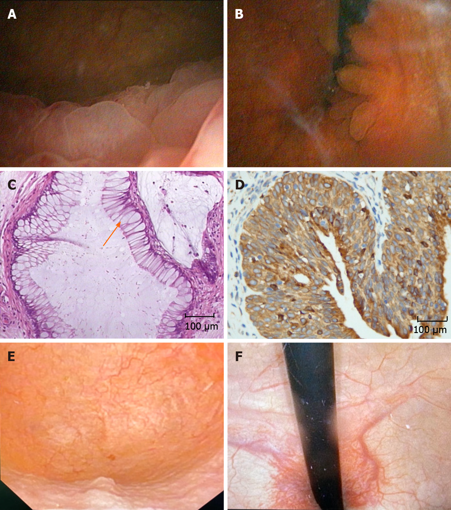
Figure 2 Cystoscopy and pathological results.
A and B: Bladder lesions in the trigonum vesicae, cervix vesicae (A), and both sides of ureteral orifices (B) were observed at the first visit; C: The initial histopathological analysis after transurethral resection of bladder tumour showed considerable goblet cells (shown by arrows) in the bladder epithelium; D: Immunohistochemical analysis of cystitis glandularis showed positive expression of cyclooxygenase-2 (COX-2) in the cytoplasm. The pathological results showed that it was intestinal glandular cystitis; E and F: Negative recurrence of bladder tumor was observed cystoscopically after 6-mo application of COX-2 inhibitor.
- Citation: Mo LC, Piao SZ, Zheng HH, Hong T, Feng Q, Ke M. Pelvic lipomatosis with cystitis glandularis managed with cyclooxygenase-2 inhibitor: A case report. World J Clin Cases 2021; 9(17): 4373-4380
- URL: https://www.wjgnet.com/2307-8960/full/v9/i17/4373.htm
- DOI: https://dx.doi.org/10.12998/wjcc.v9.i17.4373


