Copyright
©The Author(s) 2021.
World J Clin Cases. Jun 16, 2021; 9(17): 4357-4364
Published online Jun 16, 2021. doi: 10.12998/wjcc.v9.i17.4357
Published online Jun 16, 2021. doi: 10.12998/wjcc.v9.i17.4357
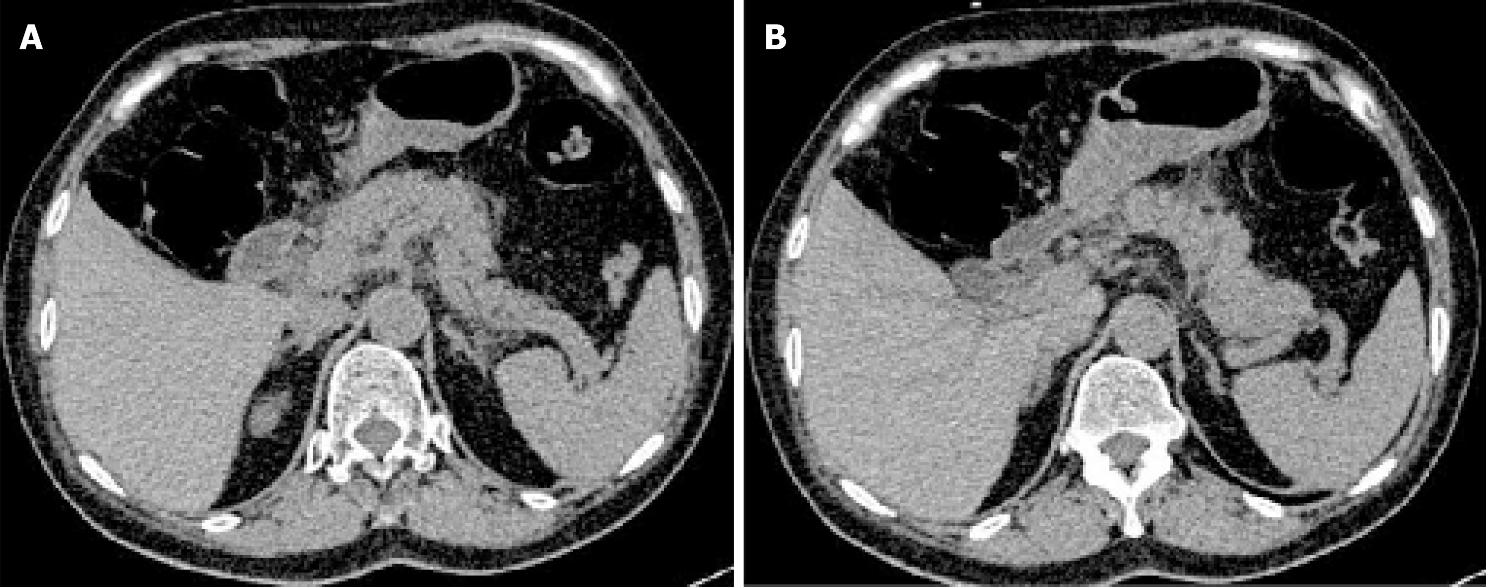
Figure 1 Abdominal computed tomography (first admission) showing pancreatic edema and peripancreatic exudation, which are characteristic findings of acute pancreatitis.
A: Edema and peripancreatic exudation in head and body of the pancreas; B: Swelling in body and tail of the pancreas.
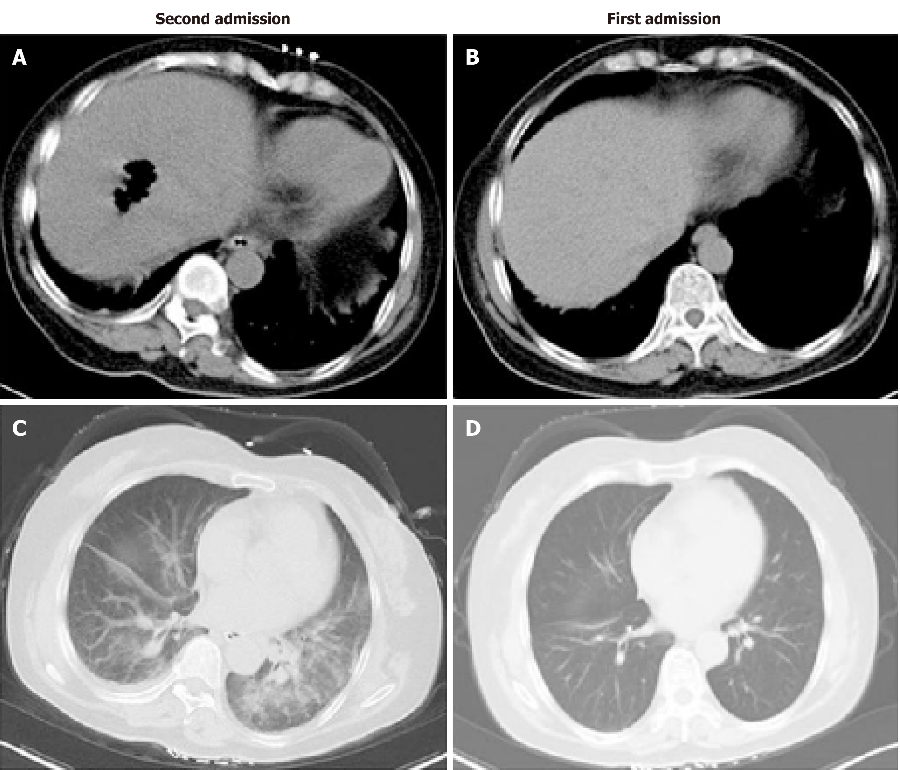
Figure 2 Computed tomography findings.
There were about 22 h between the two scans. A: Abdominal computed tomography (second admission) showing a cavity with gas in the liver; B: Abdominal computed tomography (first admission) showing no abnormal findings in the liver; C: Chest computed tomography (second admission) showing exudation of the two lungs; D: Chest computed tomography (first admission) showing no exudation.
- Citation: Li M, Li N. Clostridium perfringens bloodstream infection secondary to acute pancreatitis: A case report. World J Clin Cases 2021; 9(17): 4357-4364
- URL: https://www.wjgnet.com/2307-8960/full/v9/i17/4357.htm
- DOI: https://dx.doi.org/10.12998/wjcc.v9.i17.4357


