Copyright
©The Author(s) 2021.
World J Clin Cases. Jun 16, 2021; 9(17): 4348-4356
Published online Jun 16, 2021. doi: 10.12998/wjcc.v9.i17.4348
Published online Jun 16, 2021. doi: 10.12998/wjcc.v9.i17.4348
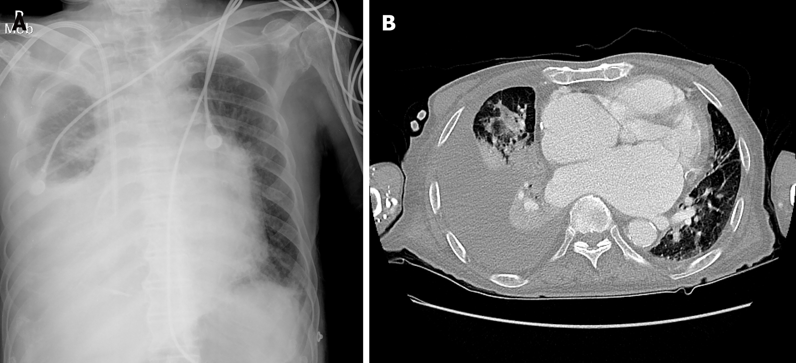
Figure 1 Radiographic imaging.
A: Chest radiograph showing a large right pleural effusion and pulmonary edema; B: Chest computed tomography showing consolidation of the right middle lobe, total atelectasis of the right lower lobe, and a large right pleural effusion.
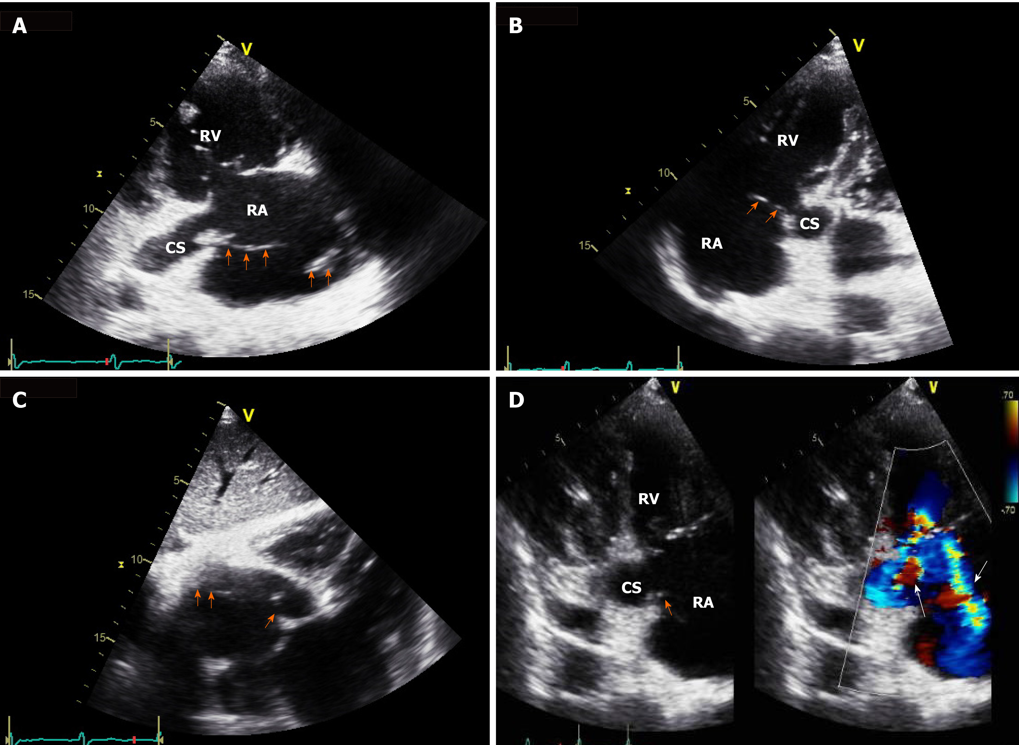
Figure 2 Echocardiographic imaging.
A: Right ventricular inflow view showing a mobile band-like vegetation, approximately 8 cm in size, attached to the coronary sinus ostium and the posterolateral wall of the right atrium; B: Modified apical four-chamber view showing a vegetation attached to the ostium of the coronary sinus; C: Subcostal view showing a vegetation; D: Right ventricular inflow view showing eccentric tricuspid regurgitant jet flow directed towards the coronary sinus and concentric tricuspid regurgitant jet flow directed towards the posterolateral wall of the right atrium, observed through color Doppler imaging, and an attached vegetation at the site, observed via two-dimensional imaging. Orange arrow indicates vegetation; White arrow indicates directed tricuspid regurgitant jet flow. RA: Right atrium; RV: Right ventricle; CS: Coronary sinus.
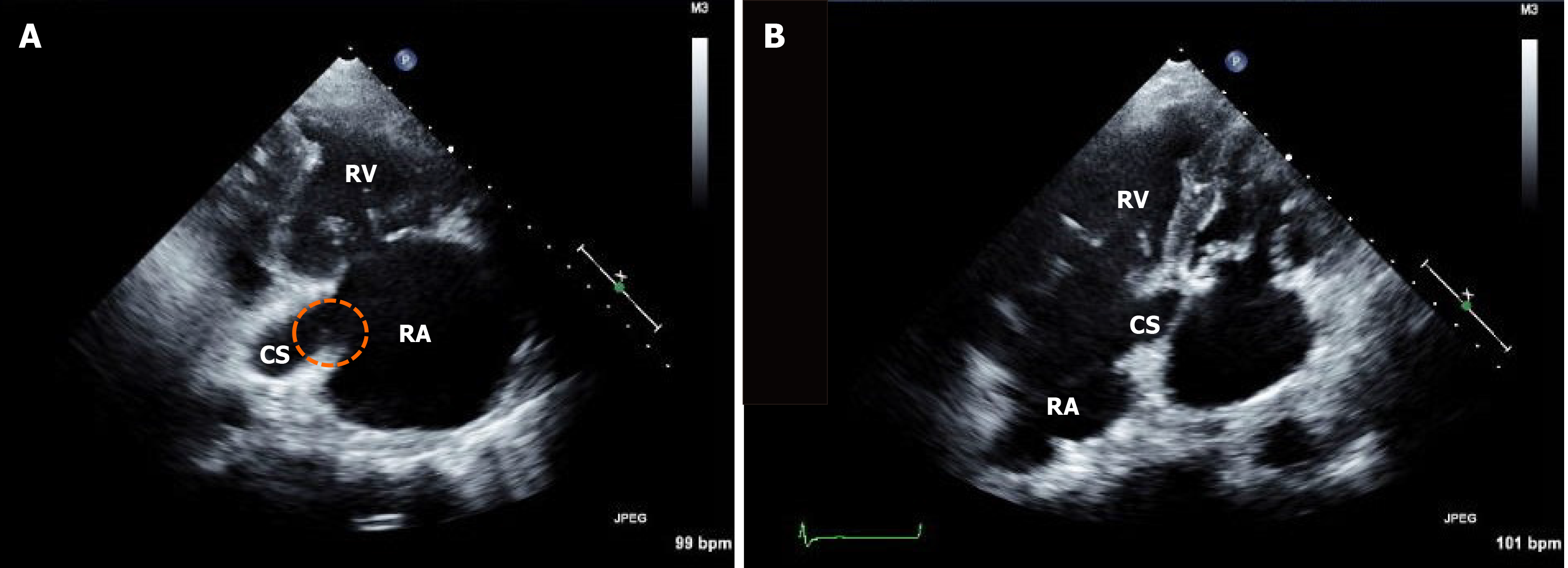
Figure 3 Follow-up echocardiographic imaging.
A: Right ventricular inflow; B: Modified apical four-chamber views showing a remnant vegetation at the coronary sinus ostium (dotted circle). RA: Right atrium; RV: Right ventricle; CS: Coronary sinus.
- Citation: Hwang HJ, Kang SW. Coronary sinus endocarditis in a hemodialysis patient: A case report and review of literature. World J Clin Cases 2021; 9(17): 4348-4356
- URL: https://www.wjgnet.com/2307-8960/full/v9/i17/4348.htm
- DOI: https://dx.doi.org/10.12998/wjcc.v9.i17.4348


