Copyright
©The Author(s) 2021.
World J Clin Cases. Jun 16, 2021; 9(17): 4342-4347
Published online Jun 16, 2021. doi: 10.12998/wjcc.v9.i17.4342
Published online Jun 16, 2021. doi: 10.12998/wjcc.v9.i17.4342
Figure 1 Preoperative imaging examination.
A: The colonoscopy suggested ulcerative colonic lesions with a narrow lumen; B: Computed tomography indicated increased liver and spleen volume; C: Computed tomography indicated changes in the left transverse colon wall and significant narrowing of the intestinal lumen, leading to proximal colonic obstruction and fecal accumulation.
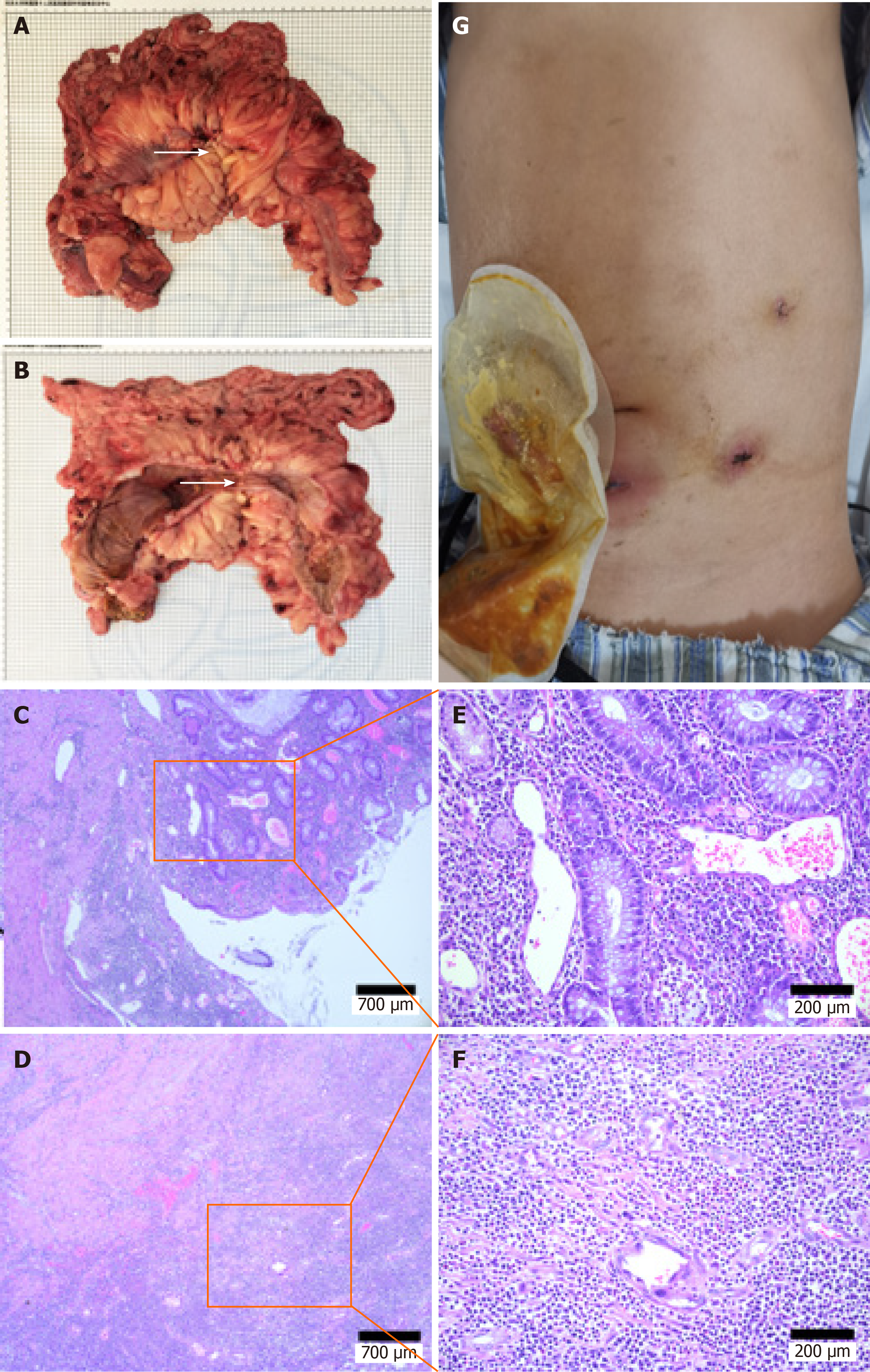
Figure 2 Photograph of the specimen and histologic features of the resected colon.
A and B: Partial transverse colon was thickened and narrowed, (white arrow); C-F: Postoperative pathologic evaluation revealed chronic ulcer and intestinal abscess formation in the colon; G: Patient’s abdomen after surgery.
- Citation: Wan J, Zhang ZC, Yang MQ, Sun XM, Yin L, Chen CQ. Minimally invasive surgery for glycogen storage disease combined with inflammatory bowel disease: A case report. World J Clin Cases 2021; 9(17): 4342-4347
- URL: https://www.wjgnet.com/2307-8960/full/v9/i17/4342.htm
- DOI: https://dx.doi.org/10.12998/wjcc.v9.i17.4342


