Copyright
©The Author(s) 2021.
World J Clin Cases. Jun 16, 2021; 9(17): 4303-4309
Published online Jun 16, 2021. doi: 10.12998/wjcc.v9.i17.4303
Published online Jun 16, 2021. doi: 10.12998/wjcc.v9.i17.4303
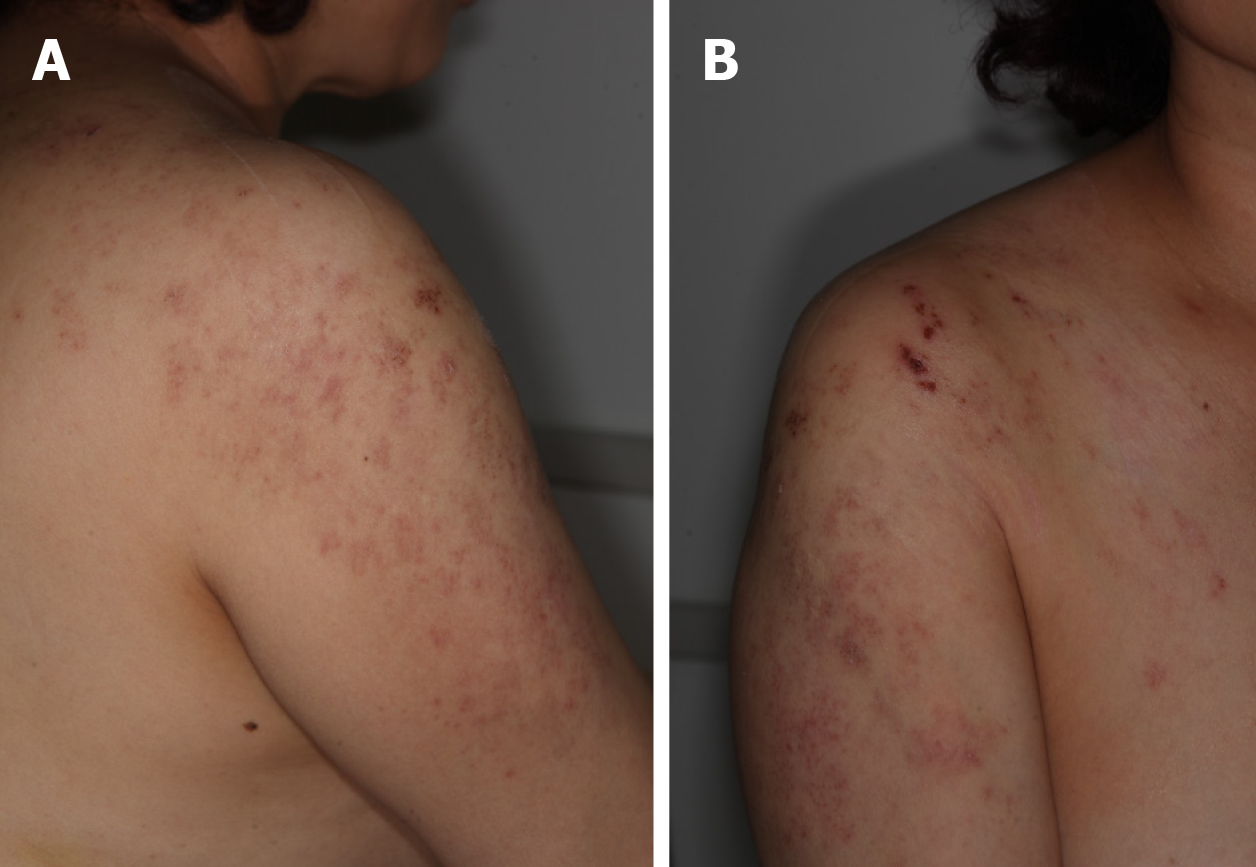
Figure 1 Multiple erythematous grouped vesicles were found on the right C4-5 and T1 dermatome regions.
A: Posterior view of the right C4-5 dermatome; B: Anterior view of the right C4-5 and T1 dermatomes.
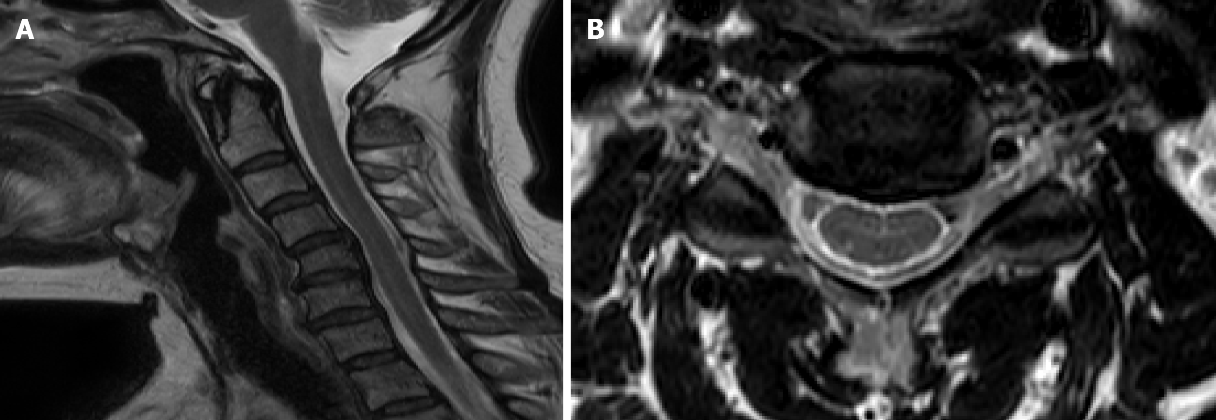
Figure 2 Magnetic resonance imaging of the cervical spine.
A: Sagittal view of magnetic resonance imaging of the cervical spine. Cervical kyphosis and spondylosis with spur formation were noted at C4-C6; B: Axial view of magnetic resonance imaging of the cervical spine, between C4 and C5. Mild intervertebral disc herniation was noted at C4-C5 without evidence of nerve root compression.
- Citation: Kim HS, Jung JW, Jung YJ, Ro YS, Park SB, Lee KH. Complete recovery of herpes zoster radiculopathy based on electrodiagnostic study: A case report. World J Clin Cases 2021; 9(17): 4303-4309
- URL: https://www.wjgnet.com/2307-8960/full/v9/i17/4303.htm
- DOI: https://dx.doi.org/10.12998/wjcc.v9.i17.4303


