Copyright
©The Author(s) 2021.
World J Clin Cases. Jun 16, 2021; 9(17): 4294-4302
Published online Jun 16, 2021. doi: 10.12998/wjcc.v9.i17.4294
Published online Jun 16, 2021. doi: 10.12998/wjcc.v9.i17.4294
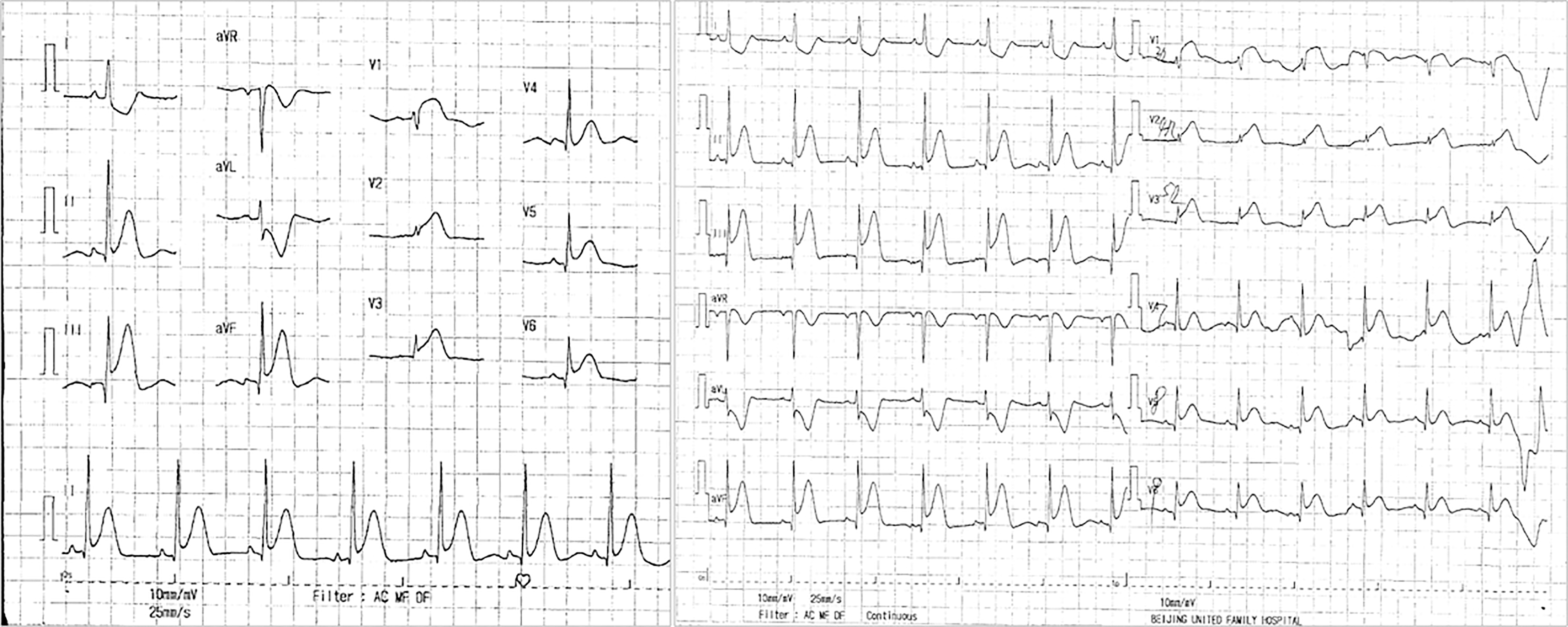
Figure 1 Eighteen-lead electrocardiogram showed ST-segment elevation in leads II, III, aVF, V3R-V5R, and V7-V9.
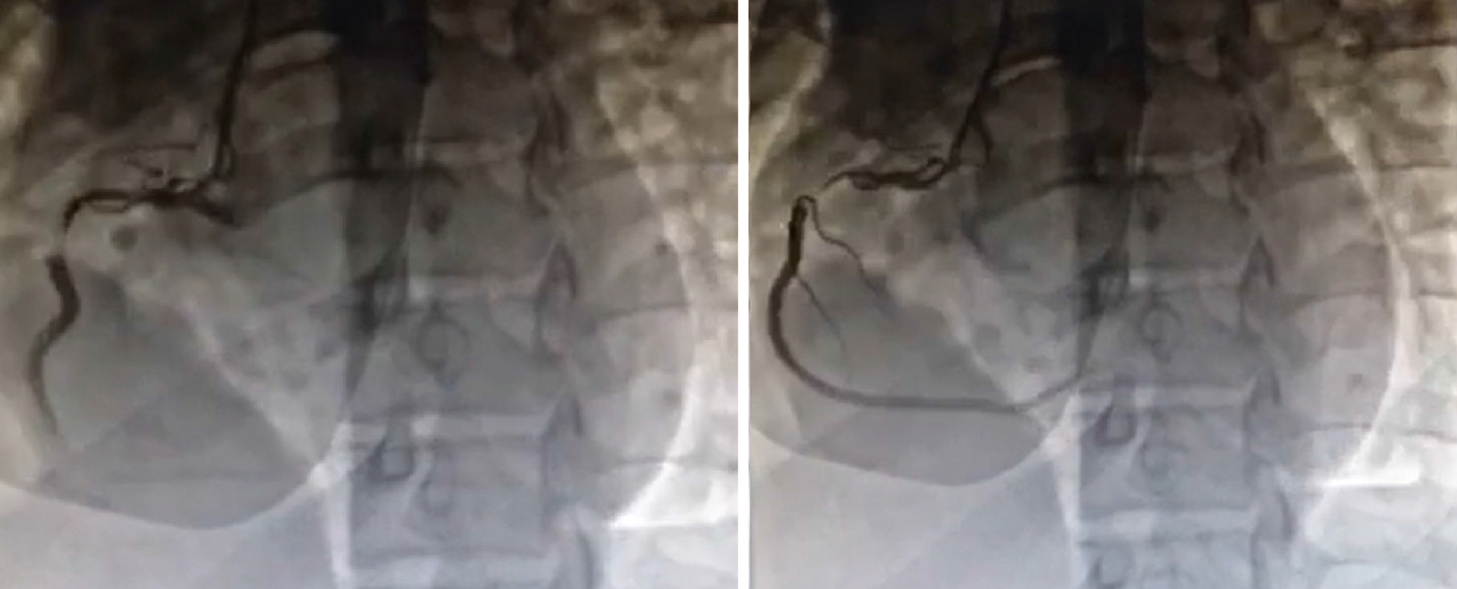
Figure 2 Right coronary angiography showed that the proximal segment of the right coronary artery had the most severe stenosis (90%) and thrombus shadow; thrombolysis in myocardial infarction flow grade 3 was observed.
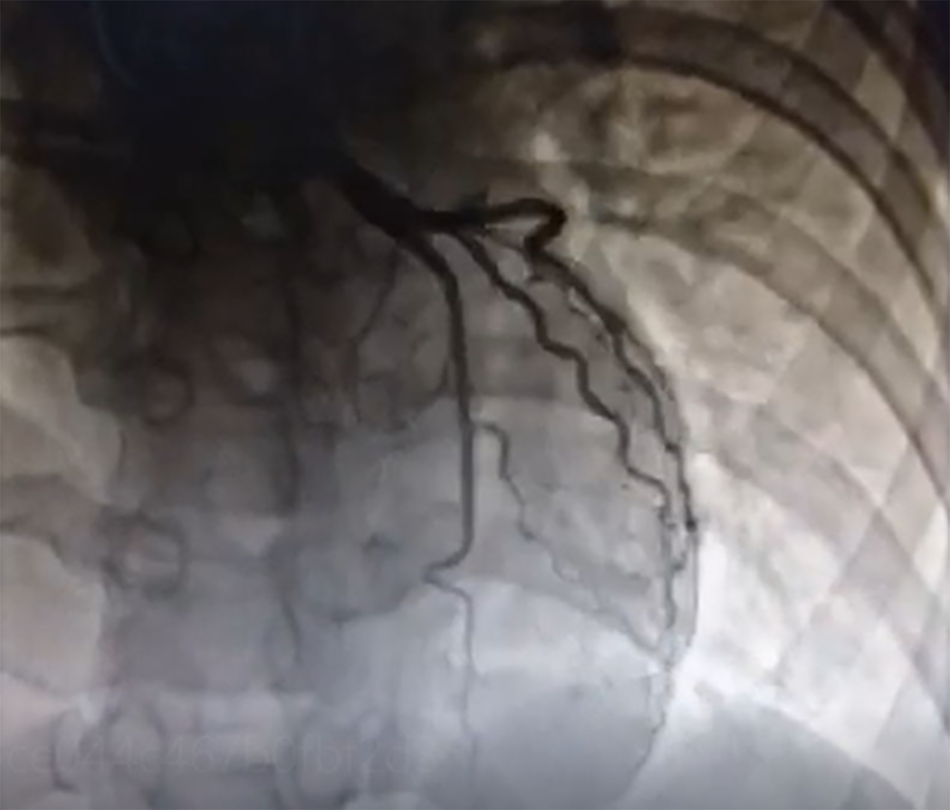
Figure 3 Left coronary angiography showed no obvious abnormalities.
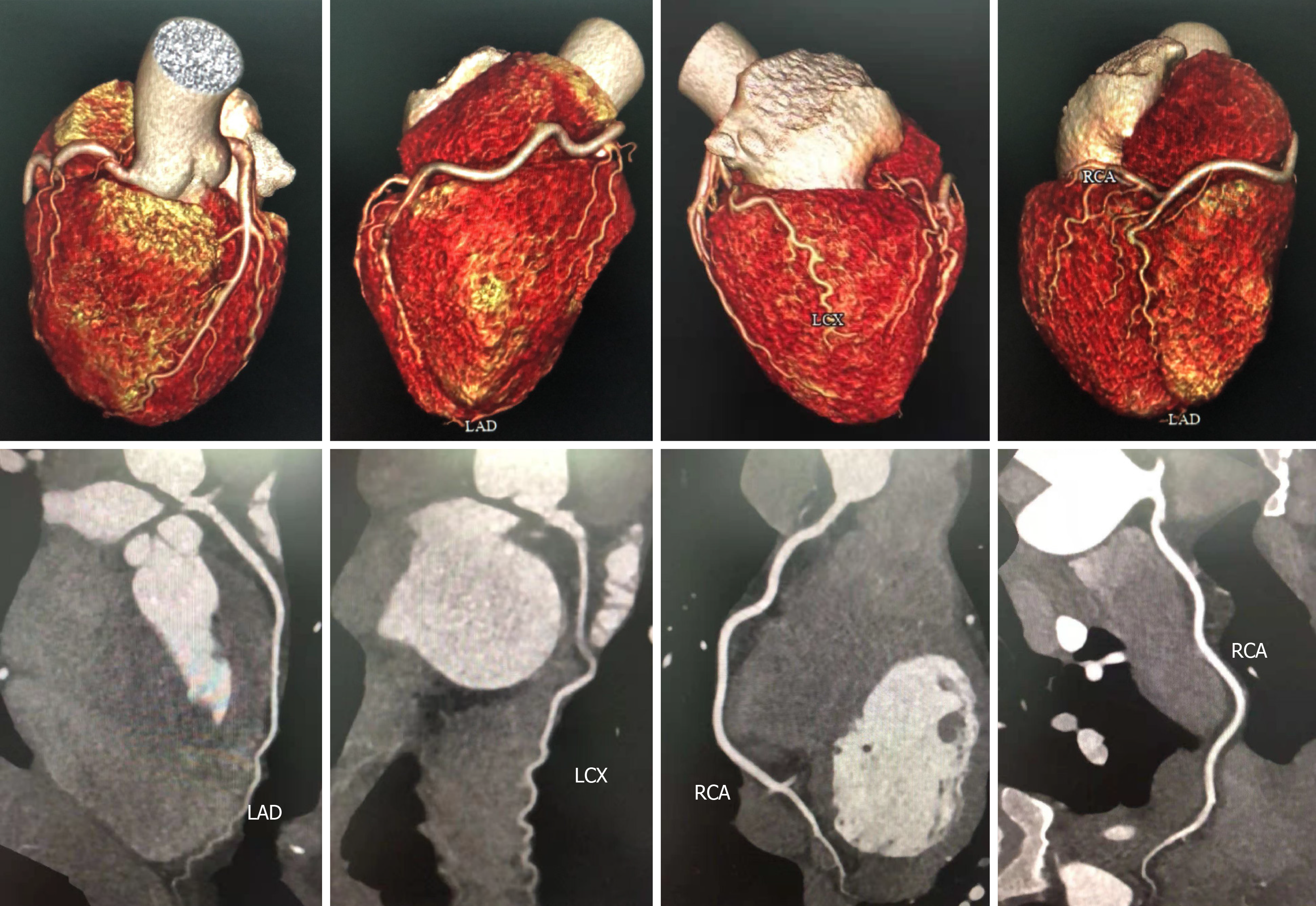
Figure 4 Coronary computed tomography angioplasty showed that there was no significant stenosis in the left main coronary artery, left circumflex branch, and right coronary artery.
The middle segment of the left anterior descending branch had mild stenosis (< 50%). LAD: Left anterior descending coronary artery; LCX: Left circumflex branch; RCA: Right coronary artery.
- Citation: Dai NN, Zhou R, Zhuo YL, Sun L, Xiao MY, Wu SJ, Yu HX, Li QY. Acute myocardial infarction in twin pregnancy after assisted reproduction: A case report. World J Clin Cases 2021; 9(17): 4294-4302
- URL: https://www.wjgnet.com/2307-8960/full/v9/i17/4294.htm
- DOI: https://dx.doi.org/10.12998/wjcc.v9.i17.4294


