Copyright
©The Author(s) 2021.
World J Clin Cases. Jun 16, 2021; 9(17): 4268-4278
Published online Jun 16, 2021. doi: 10.12998/wjcc.v9.i17.4268
Published online Jun 16, 2021. doi: 10.12998/wjcc.v9.i17.4268
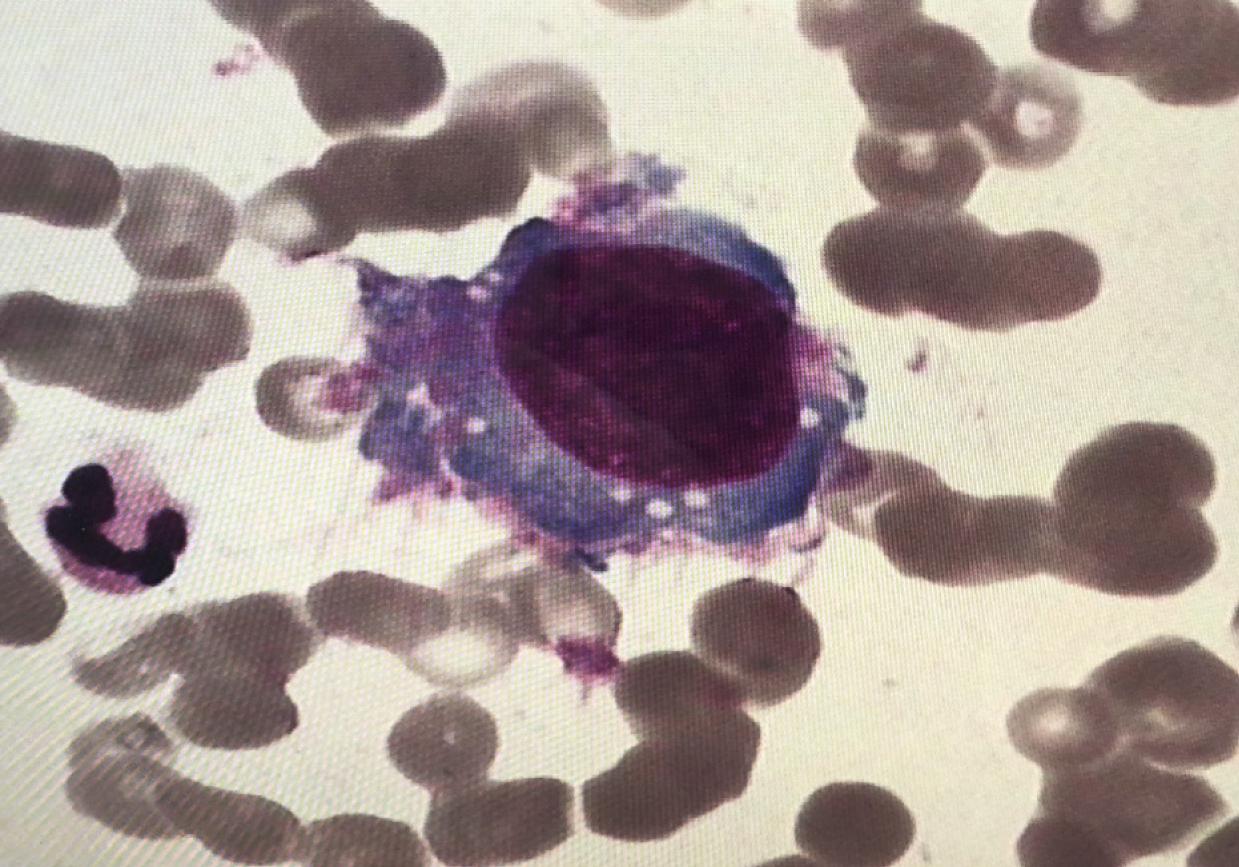
Figure 1 Tumor cells found in bone marrow smears (Wright-Giemsa-stained, 1000 ×).
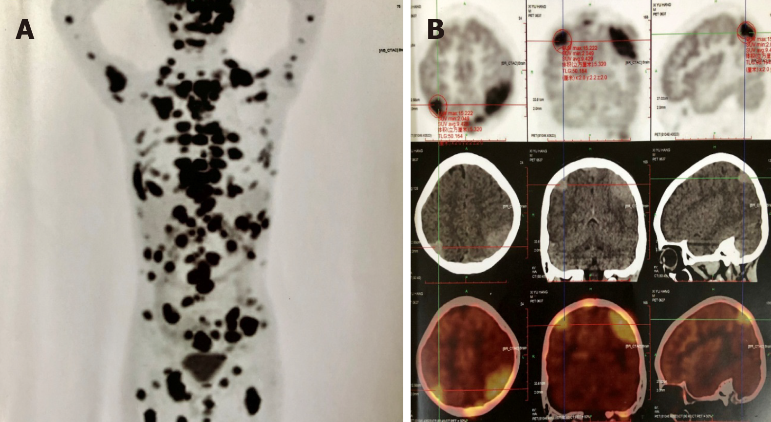
Figure 2 Positron emission tomography/computed tomography scan.
A: Trunk; B: Cranium.
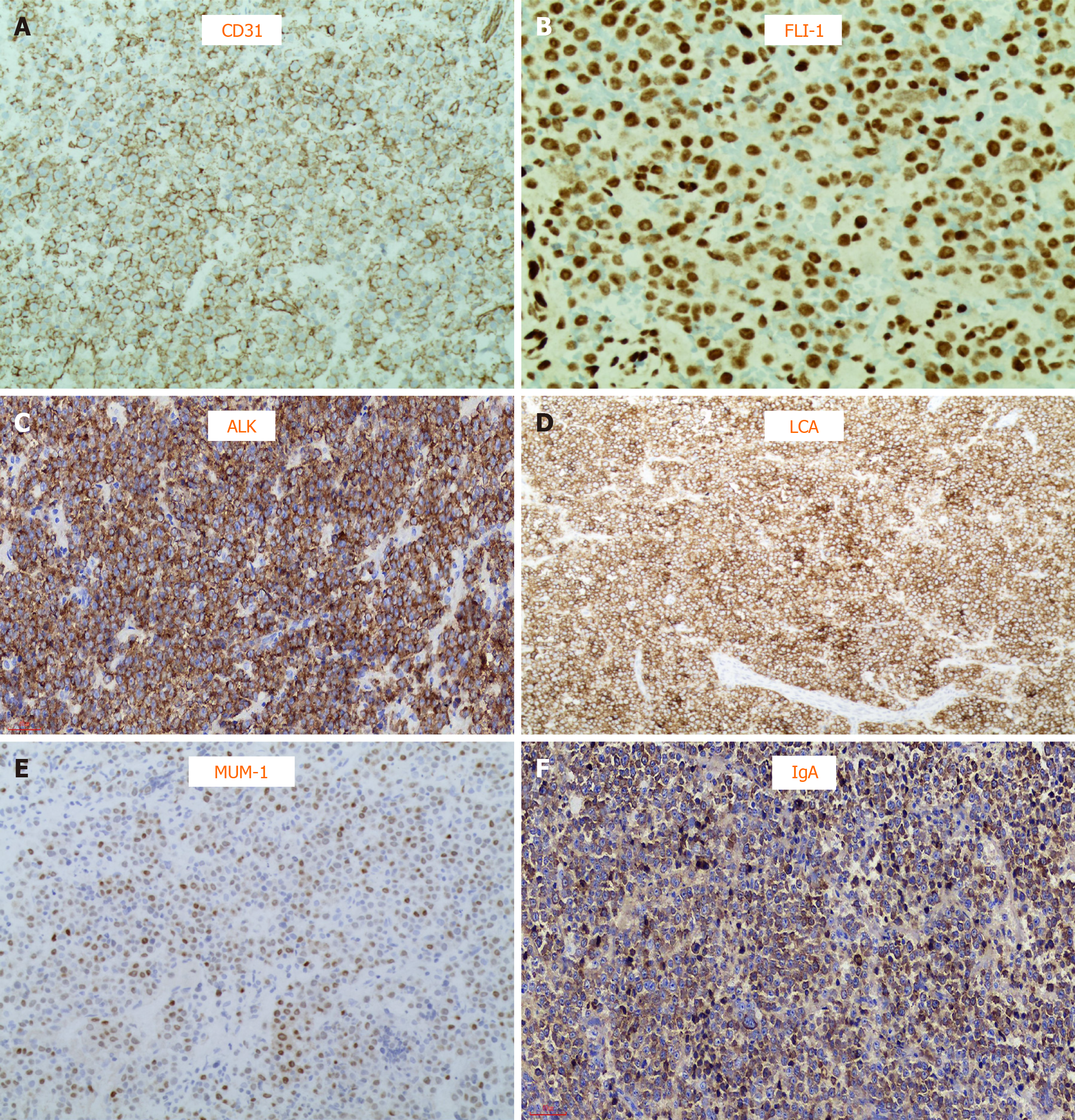
Figure 3 Hematoxylin and eosin-stained section of neck lymph node biopsy (200 ×).
A: Positive cluster of differentiation 31 staining; B: Positive friend leukemia integration 1 staining; C: Positive anaplastic lymphoma kinase staining; D: Positive leukocyte-common antigen staining; E: Positive multiple myeloma oncogene 1 staining; F: Positive immunoglobulin A staining.
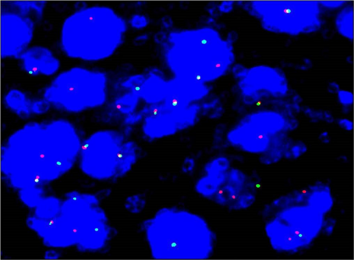
Figure 4 Fluorescence in situ hybridization study with ALK breakapart probe showed ALK gene disruption.
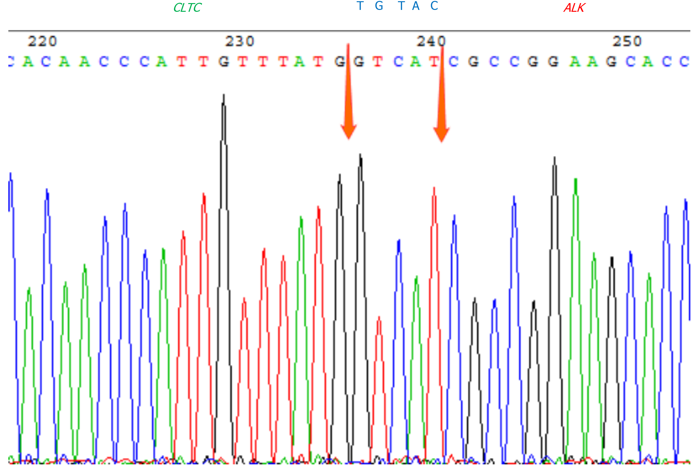
Figure 5 CLTC-ALK fusion-positive in bone marrow.
- Citation: Zhang M, Jin L, Duan YL, Yang J, Huang S, Jin M, Zhu GH, Gao C, Liu Y, Zhang N, Zhou CJ, Gao ZF, Zheng QL, Chen D, Zhang YH. Diagnosis and treatment of pediatric anaplastic lymphoma kinase-positive large B-cell lymphoma: A case report. World J Clin Cases 2021; 9(17): 4268-4278
- URL: https://www.wjgnet.com/2307-8960/full/v9/i17/4268.htm
- DOI: https://dx.doi.org/10.12998/wjcc.v9.i17.4268


