Copyright
©The Author(s) 2021.
World J Clin Cases. Jun 16, 2021; 9(17): 4253-4261
Published online Jun 16, 2021. doi: 10.12998/wjcc.v9.i17.4253
Published online Jun 16, 2021. doi: 10.12998/wjcc.v9.i17.4253
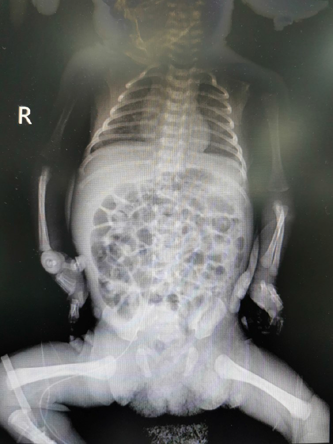
Figure 1
The tip position was in the 8th thoracic vertebra.
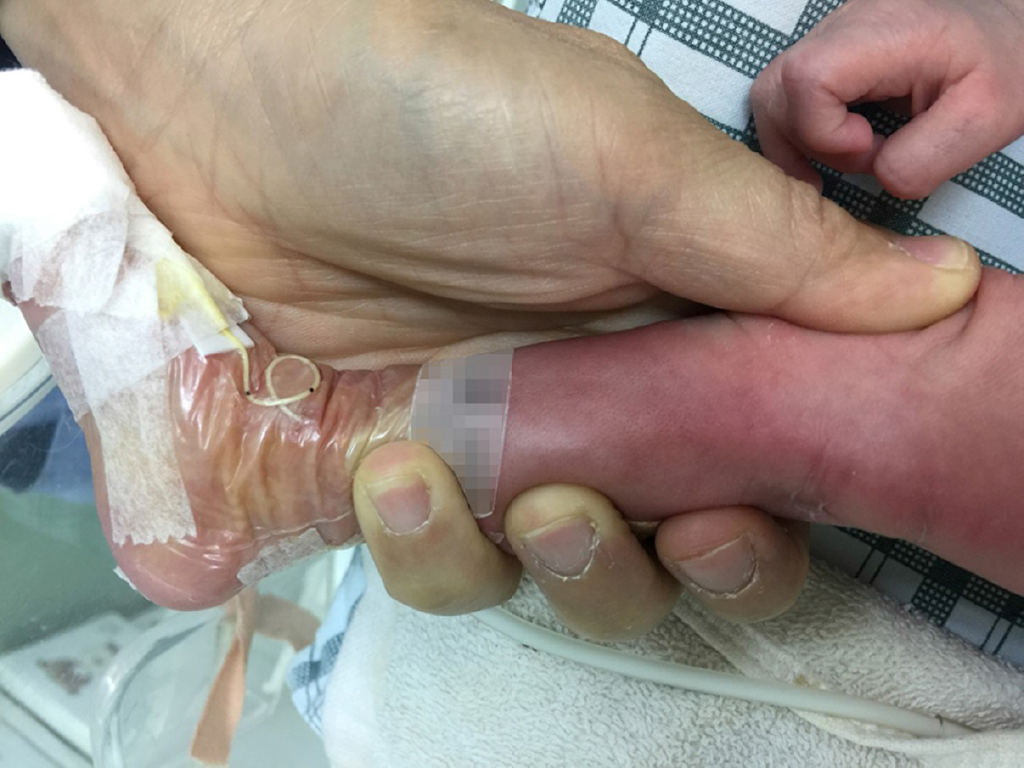
Figure 2
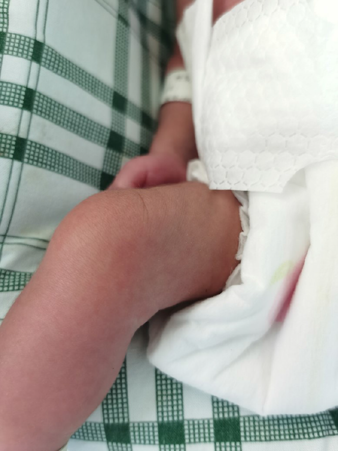
The local skin redness occurred around the puncture site 22 h after catheterization.
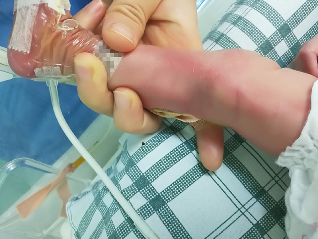
Figure 3
The area of redness and right leg circumference increased on the second day of catheterization.
Figure 4
Cordage presented on the puncture point on the third day of catheterization.
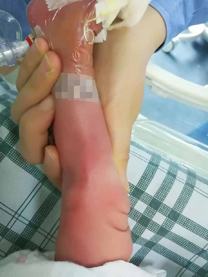
Figure 5
The red swelling increased more on the fourth day of catheterization.
Figure 6 Imaging examinations.
A: The tip position was in the 10th thoracic vertebra on the day of peripherally inserted central catheter removal; B: The state of the veins and the surrounding tissue at the first ultrasound; C: The state of the veins and the surrounding tissue at the second ultrasound.
Figure 7
Recovery from phlebitis after extubation.
- Citation: Chen Q, Hu YL, Su SY, Huang X, Li YX. “AFGP” bundles for an extremely preterm infant who underwent difficult removal of a peripherally inserted central catheter: A case report. World J Clin Cases 2021; 9(17): 4253-4261
- URL: https://www.wjgnet.com/2307-8960/full/v9/i17/4253.htm
- DOI: https://dx.doi.org/10.12998/wjcc.v9.i17.4253


