Copyright
©The Author(s) 2021.
World J Clin Cases. Jun 16, 2021; 9(17): 4244-4252
Published online Jun 16, 2021. doi: 10.12998/wjcc.v9.i17.4244
Published online Jun 16, 2021. doi: 10.12998/wjcc.v9.i17.4244
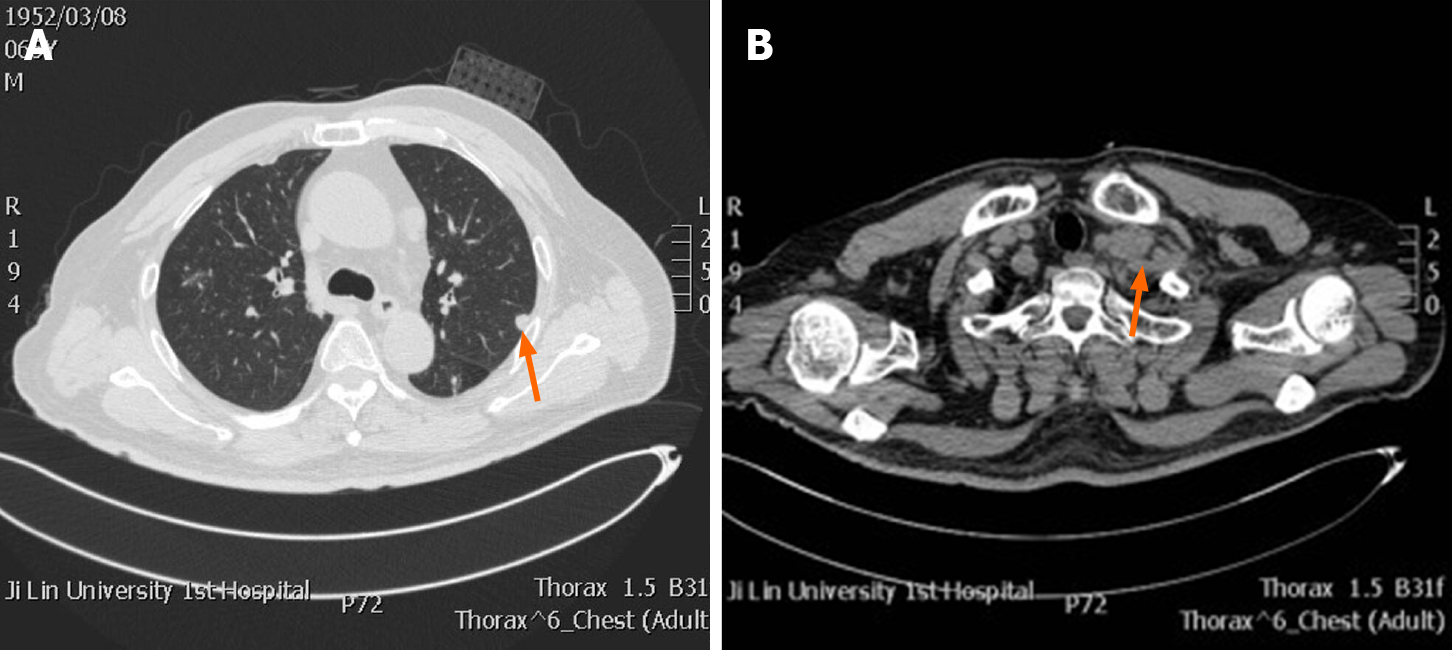
Figure 1 Chest radiologic findings.
A: A round mass was observed in the left lung lobe; B: Enlarged left supraclavicular lymph node was also noted (orange arrow indicates relevant lesions).
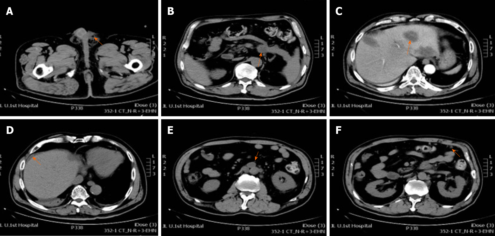
Figure 2 Postoperative abdominal radiologic findings.
A: Abdominal computed tomography shows absence of the left testis (orange arrow indicating air density spot); B: Primary tumor can be seen in the tail of pancreas; C: Multiple hepatic metastasis; D: Abdominal cavity fluid; E: Retroperitoneal lymphadenopathy; F: Omentum metastasis (orange arrow indicates relevant lesions).
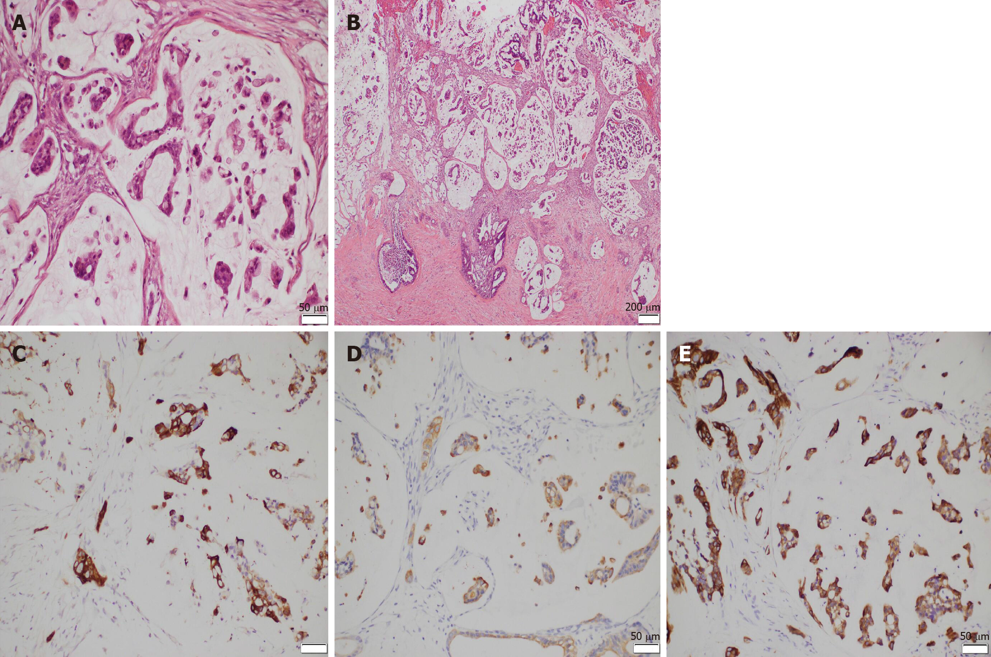
Figure 3 ematoxylin and eosin and immunohistochemical staining of the tunica vaginalis tumors.
A and B: Infiltration of malignant cells into tunica vaginalis tissue were observed (Hematoxylin and eosin, 200 ×). (A: scale = 50 μm; B: scale = 100 μm); C: Immunohistochemical staining indicates cytokeratin 5/6 (CK 5/6)-positive; D: CK20-positive; E: CK7-positive in the tunica vaginalis tumors (3, 3′ diaminobenzidine, 200×) (C, D, and E: scale = 50 μm).
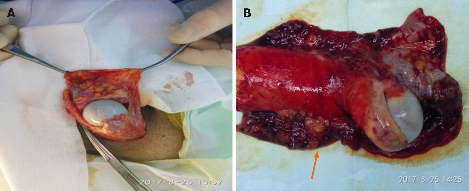
Figure 4 Gross appearance of tunica vaginalis tumors.
A: Left orchiectomy was performed via an inguinal canal approach; B: Macroscopic appearance of the surgical specimen showing multiple superficial polypoid nodules in the tunica vaginalis (orange arrow).
- Citation: Zhang YR, Ma DK, Gao BS, An W, Guo KM. Tunica vaginalis testis metastasis as the first clinical manifestation of pancreatic adenocarcinoma: A case report. World J Clin Cases 2021; 9(17): 4244-4252
- URL: https://www.wjgnet.com/2307-8960/full/v9/i17/4244.htm
- DOI: https://dx.doi.org/10.12998/wjcc.v9.i17.4244


