Copyright
©The Author(s) 2021.
World J Clin Cases. Jun 16, 2021; 9(17): 4238-4243
Published online Jun 16, 2021. doi: 10.12998/wjcc.v9.i17.4238
Published online Jun 16, 2021. doi: 10.12998/wjcc.v9.i17.4238
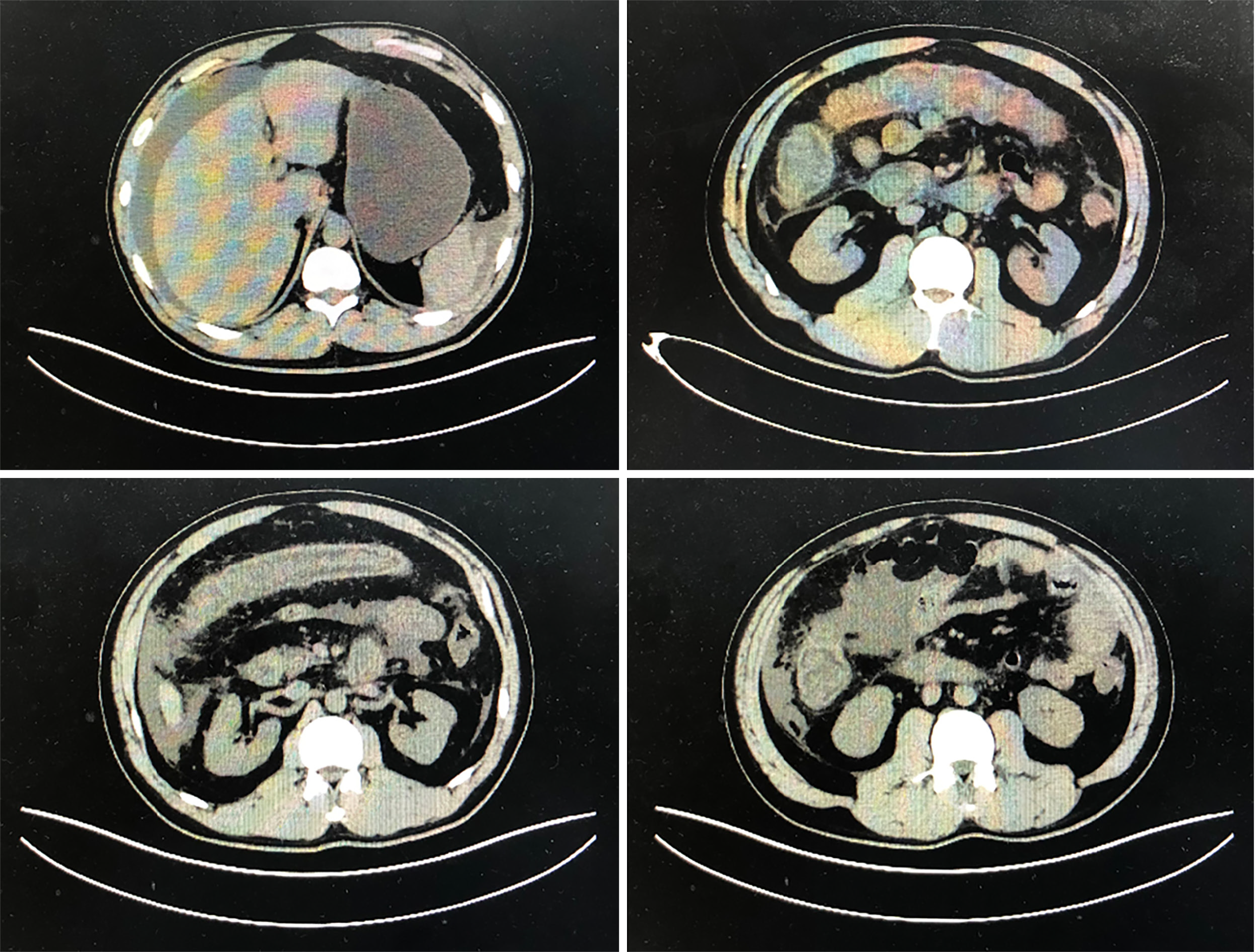
Figure 1 Abdominal computed tomography scan.
Abdominal computed tomography scan demonstrated extensive intestinal wall edema thickening in the duodenum, jejunum, ascending colon and transverse colon; multiple exudative effusion surrounding the intestinal tract; and ascites in the abdominal cavity.
Figure 2 Colonoscopy results showed the terminal ileum and entire colonic mucosa scattered in hyperemia and edema.
A: Terminal ileum; B: Transverse colon; C: Rectum.
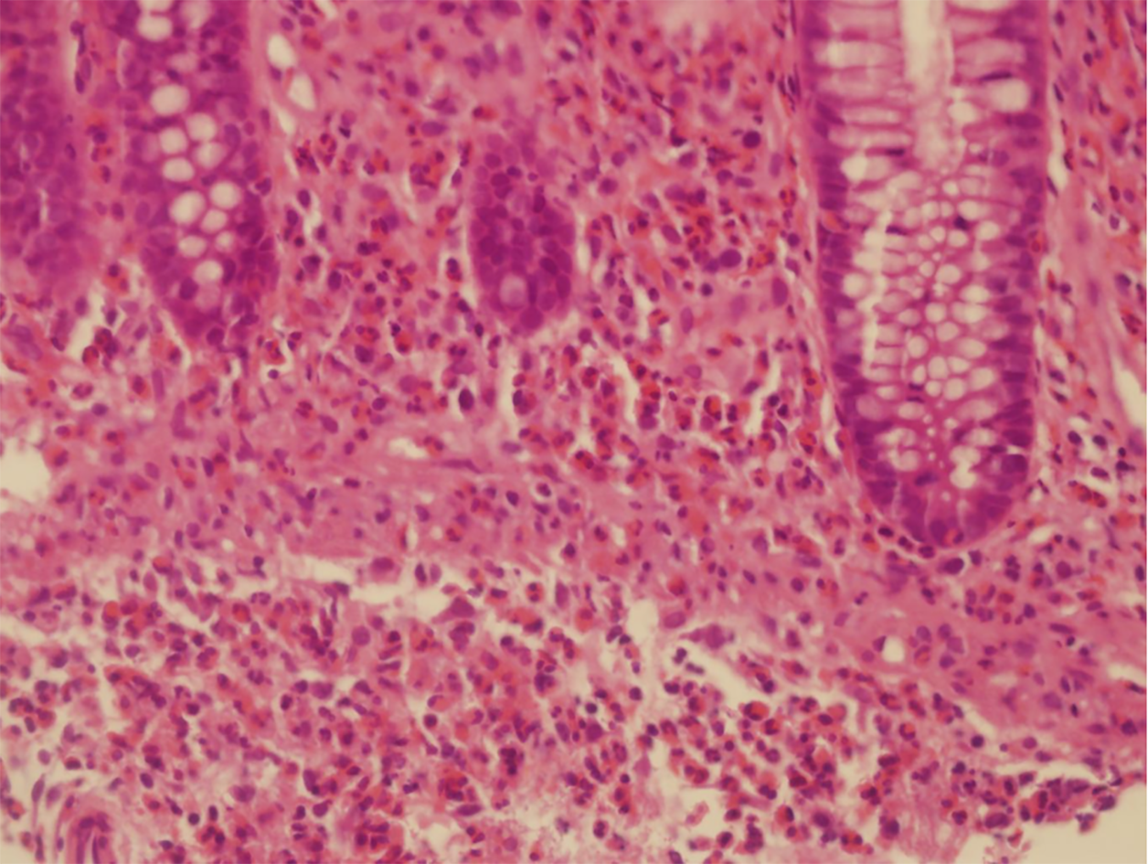
Figure 3 Biopsy results confirm the diagnosis of eosinophilic gastroenteritis.
Biopsies showed mucosa with interstitial edema and eosinophilic infiltration throughout the whole colon region with ≥ 20 eosinophils/high power field (HPF). A maximum of 50 eosinophils/HPF were found in the transverse colon (hematoxylin and eosin staining; 400 ×).
- Citation: Tian XQ, Chen X, Chen SL. Eosinophilic gastroenteritis with abdominal pain and ascites: A case report. World J Clin Cases 2021; 9(17): 4238-4243
- URL: https://www.wjgnet.com/2307-8960/full/v9/i17/4238.htm
- DOI: https://dx.doi.org/10.12998/wjcc.v9.i17.4238


