Copyright
©The Author(s) 2021.
World J Clin Cases. Jun 16, 2021; 9(17): 4166-4177
Published online Jun 16, 2021. doi: 10.12998/wjcc.v9.i17.4166
Published online Jun 16, 2021. doi: 10.12998/wjcc.v9.i17.4166
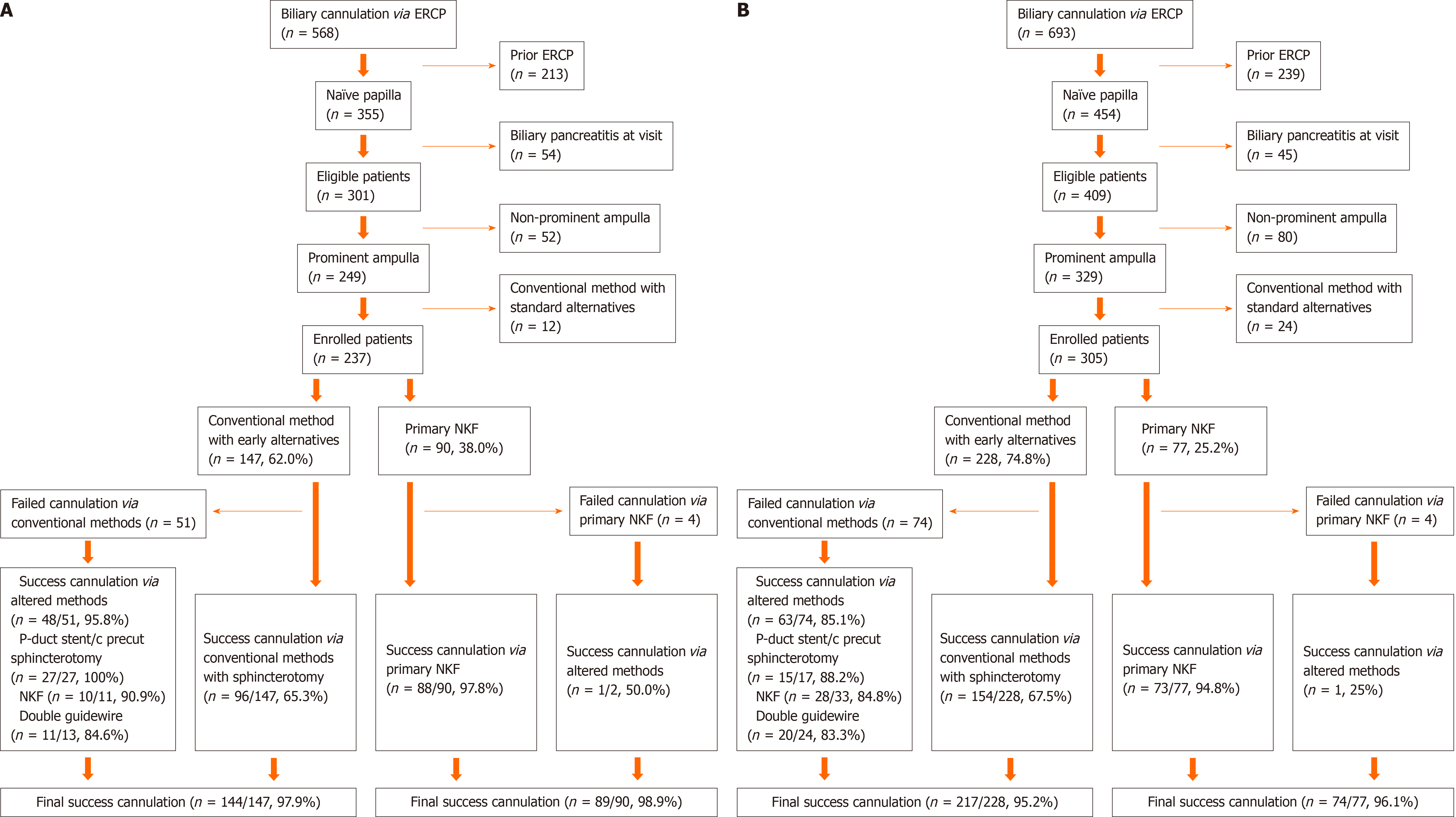
Figure 1 Study flow chart.
A: Experienced endoscopist; B: Less experienced endoscopist. ERCP: Endoscopic retrograde cholangiopancreatography; NKF: Needle-knife fistulotomy.
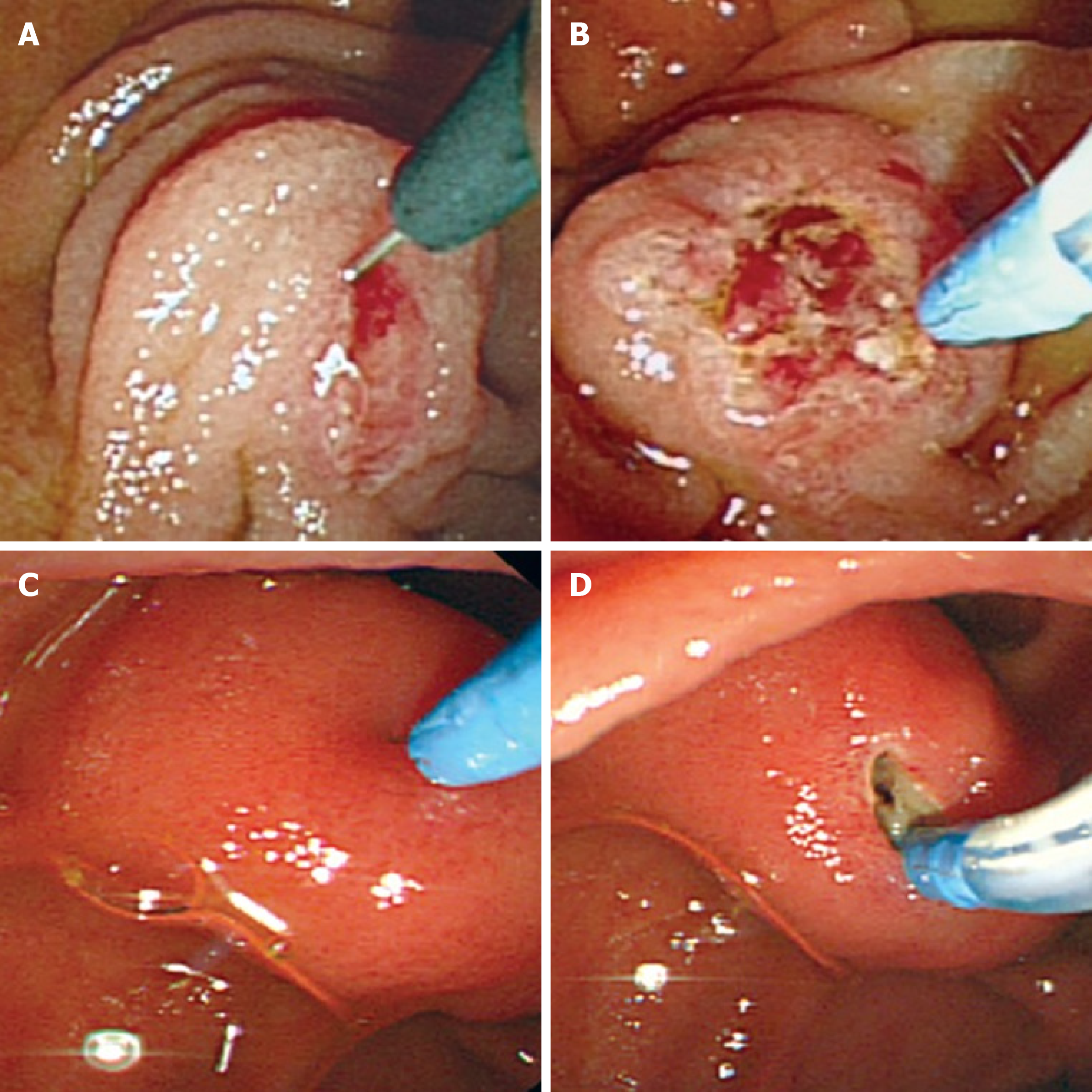
Figure 2 Comparison of precut sphincterotomy and needle-knife fistulotomy.
A: Precut sphincterotomy. The needle is placed at the orifice of the ampulla; B: Precut sphincterotomy. Precutting was performed with slight upward tension; C: Needle knife fistulotomy. Mucosal incision at the maximal bulging point of the papillary roof of the ampulla; D: Needle knife fistulotomy. Incision at the oral side of the bile duct at the 11-12 o’clock position relative to the ampulla of Vater.
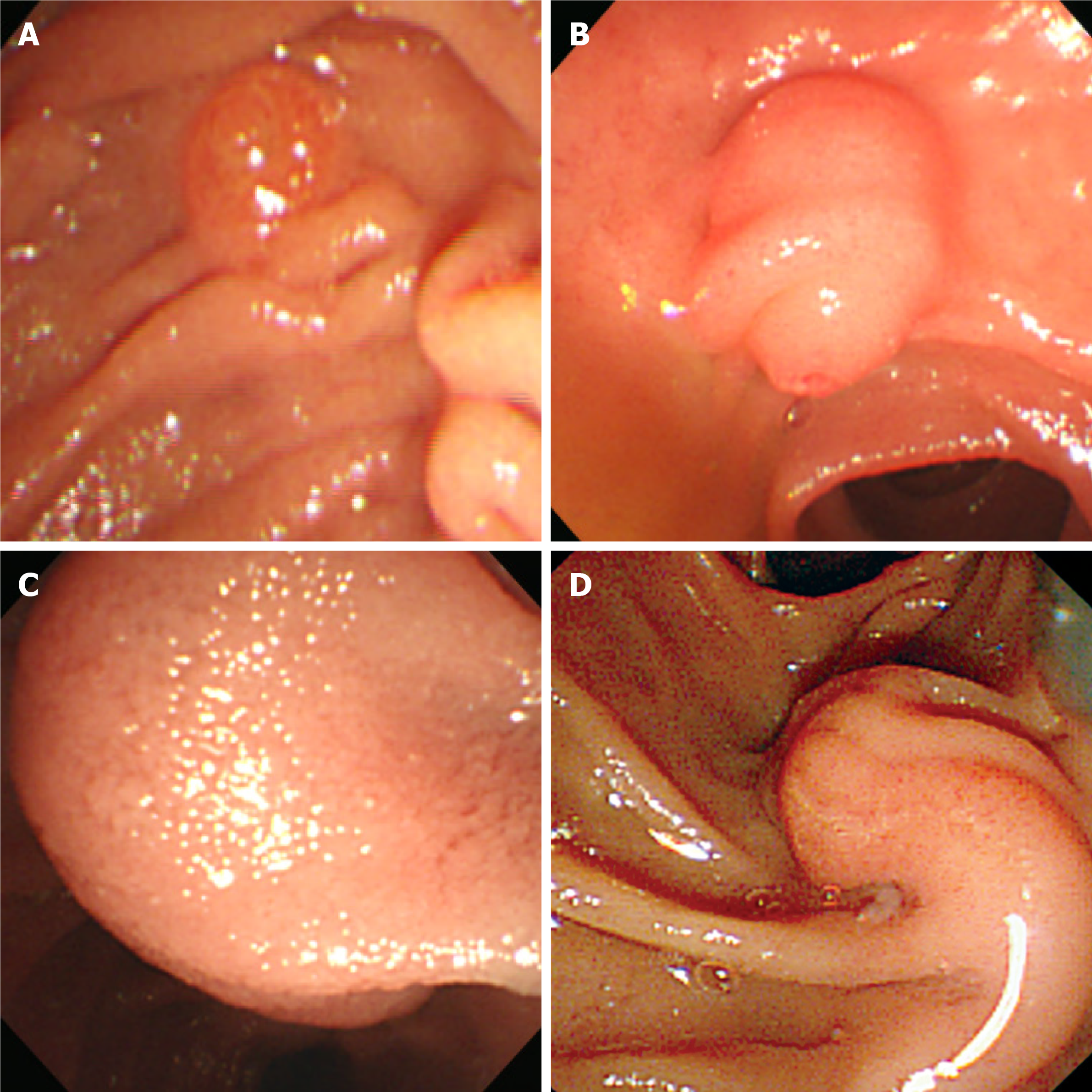
Figure 3 Ampulla configurations.
A: Non-prominent; B: Bulging; C: Hook-shape, ampulla of Vater orifice was not shown due to huge bulging; D: Distorted.
- Citation: Han SY, Baek DH, Kim DU, Park CJ, Park YJ, Lee MW, Song GA. Primary needle-knife fistulotomy for preventing post-endoscopic retrograde cholangiopancreatography pancreatitis: Importance of the endoscopist’s expertise level. World J Clin Cases 2021; 9(17): 4166-4177
- URL: https://www.wjgnet.com/2307-8960/full/v9/i17/4166.htm
- DOI: https://dx.doi.org/10.12998/wjcc.v9.i17.4166


