Copyright
©The Author(s) 2021.
World J Clin Cases. May 26, 2021; 9(15): 3779-3786
Published online May 26, 2021. doi: 10.12998/wjcc.v9.i15.3779
Published online May 26, 2021. doi: 10.12998/wjcc.v9.i15.3779
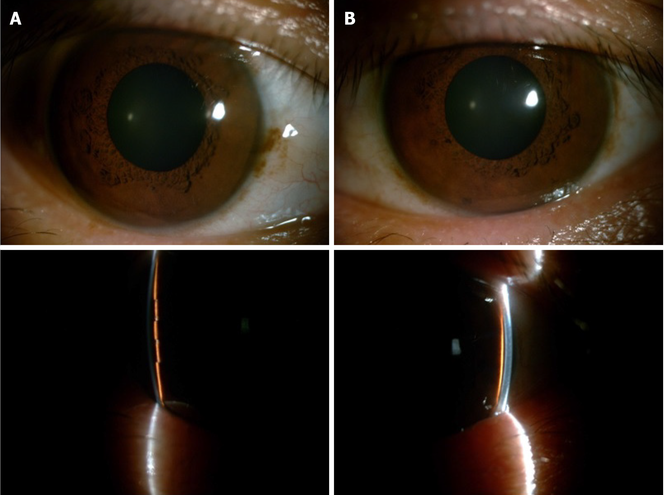
Figure 1 Anterior segment photography before treatment.
A quite shallow anterior chamber was present in both eyes and accompanied by mild cornea edema. The pupils were 5 mm, round, and symmetrical to each other. The lenses were transparent without dislocation. A: Right eye; B: Left eye.
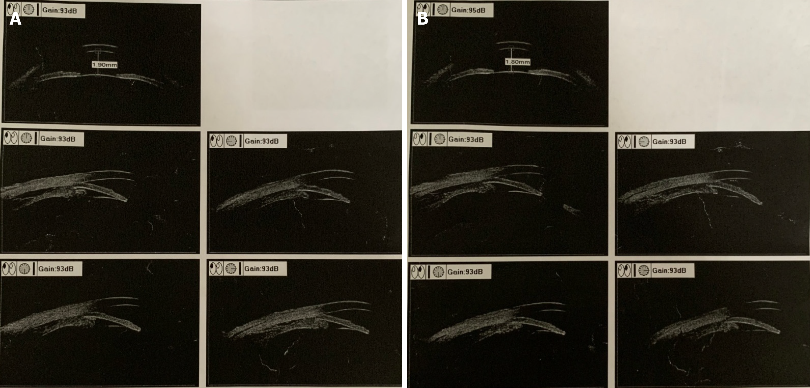
Figure 2 Ultrasound biomicroscopic examination before treatment.
The anterior chamber depths were 1.9 mm for the right eye and 1.8 mm for the left eye. Cyclodialysis, pronated ciliary process, and totally closed anterior chamber angle were present. A: Right eye; B: Left eye.
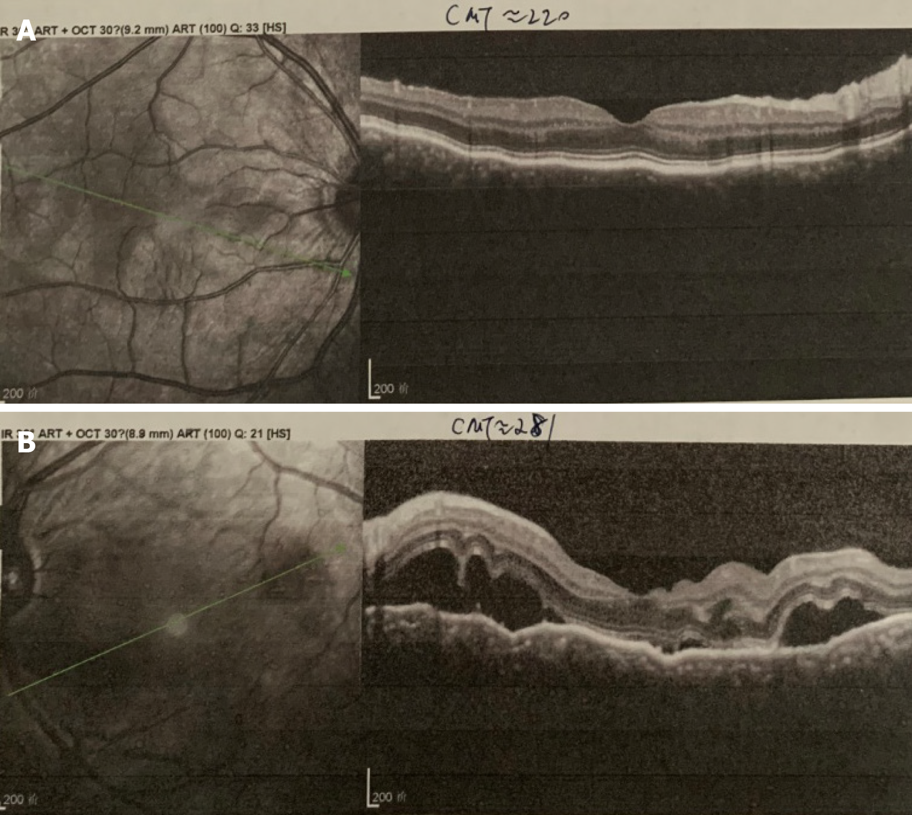
Figure 3 Optical coherence tomography examination before treatment.
A: Retinal pigment epithelial folds were found in the right eye; B: Optic disc edema and multiple serous retinal detachments were found in the left eye.
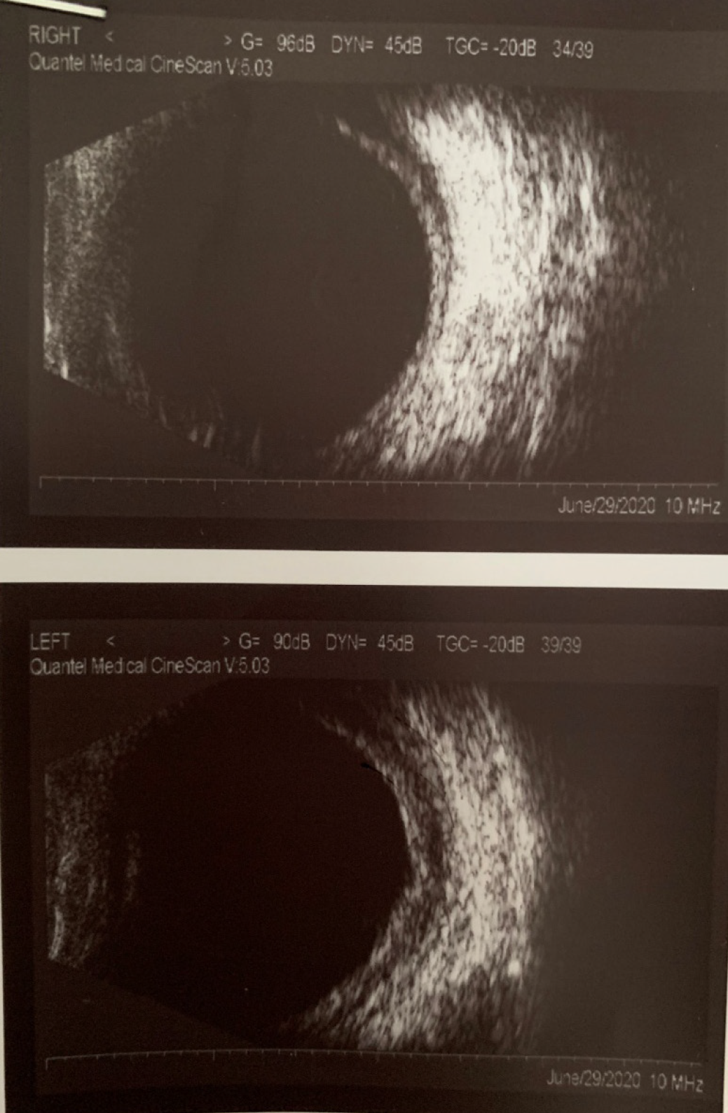
Figure 4 B-scan ultrasonography examination before treatment.
Thickening of the posterior walls was present in both eyes, as was fluid under Tenon’s capsule.
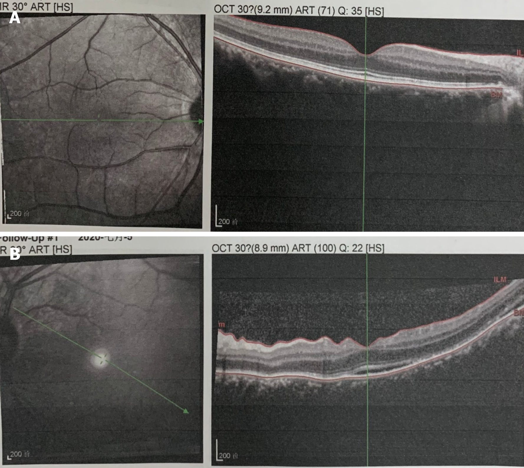
Figure 5 Optical coherence tomography examination after treatment.
Retinal pigment epithelial folds and serous detachment were found to have gradually resolved after 1 wk of treatment. A: Right eye; B: Left eye.
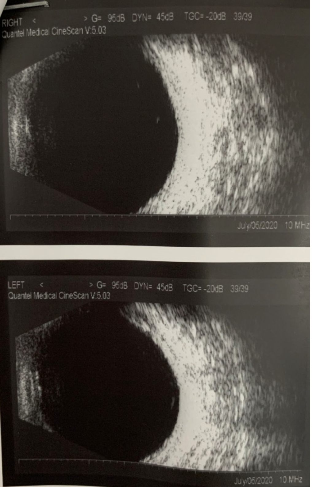
Figure 6 B-scan ultrasonography examination after treatment.
Resolution of cyclodialysis and the thinning of posterior walls were found.
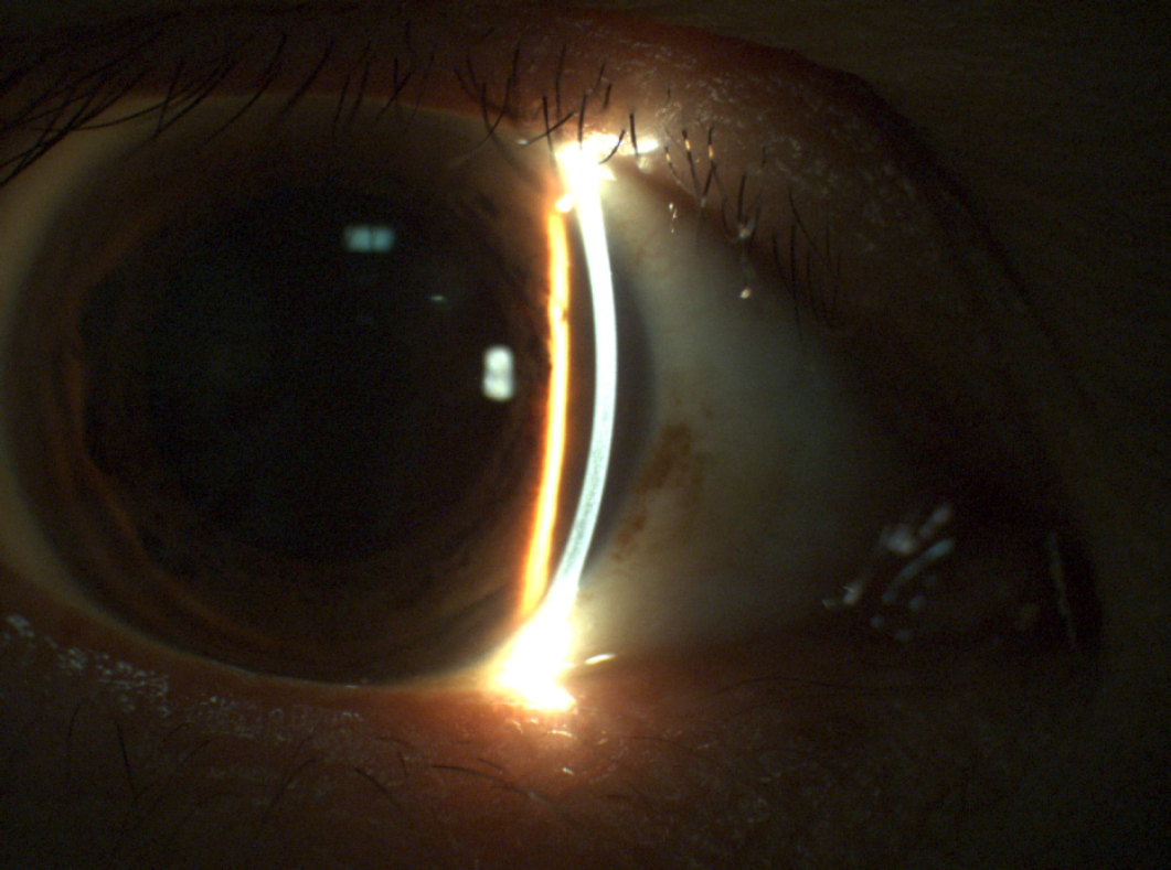
Figure 7 Slit-lamp examination after treatment.
Deepening of the anterior chamber and dilation of the pupil upon atropine sulfate eye ointment application were found.
- Citation: Wen C, Duan H. Bilateral posterior scleritis presenting as acute primary angle closure: A case report. World J Clin Cases 2021; 9(15): 3779-3786
- URL: https://www.wjgnet.com/2307-8960/full/v9/i15/3779.htm
- DOI: https://dx.doi.org/10.12998/wjcc.v9.i15.3779


