Copyright
©The Author(s) 2021.
World J Clin Cases. May 26, 2021; 9(15): 3752-3757
Published online May 26, 2021. doi: 10.12998/wjcc.v9.i15.3752
Published online May 26, 2021. doi: 10.12998/wjcc.v9.i15.3752
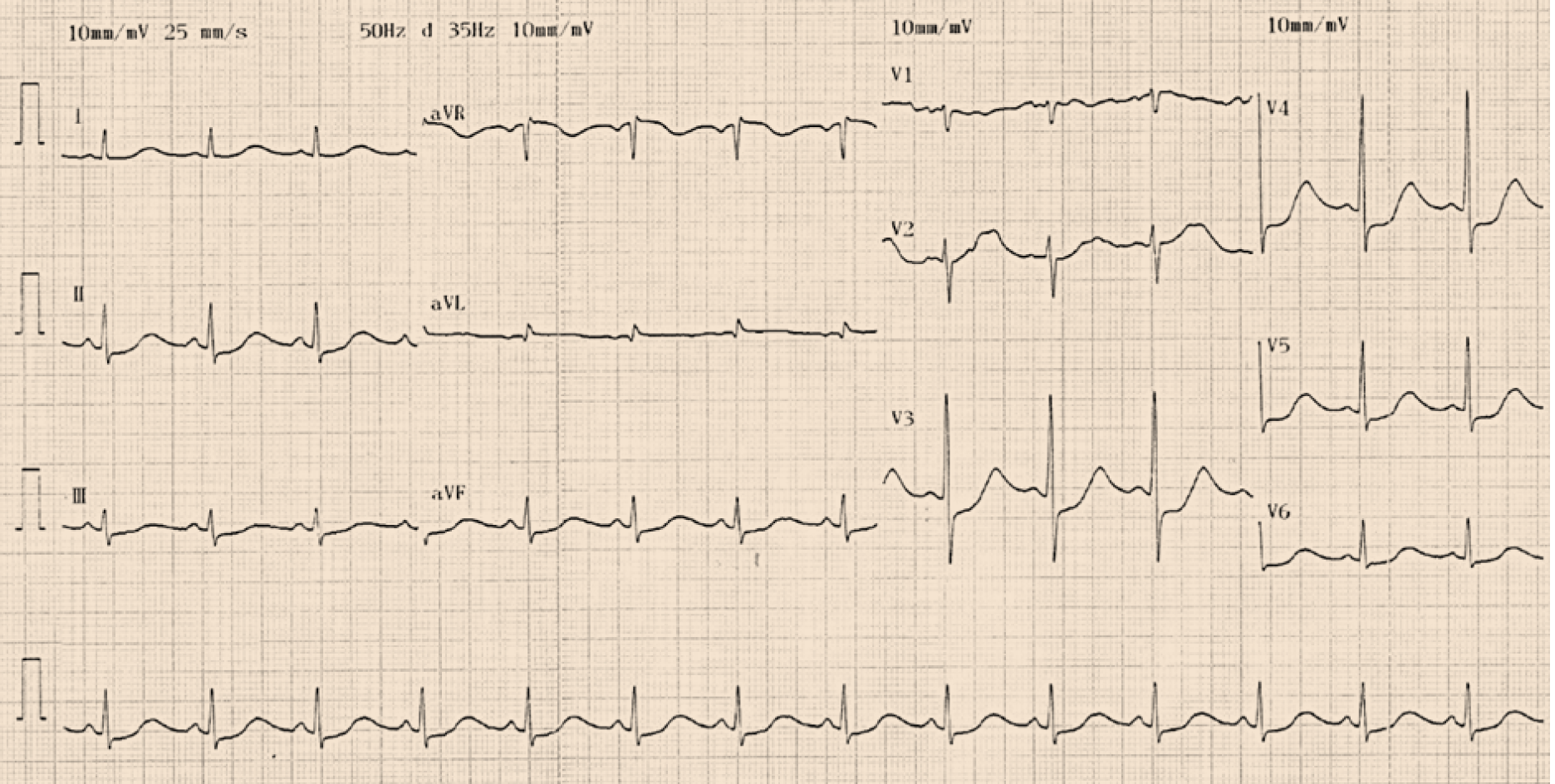
Figure 1 Twelve-lead electrocardiogram indicated QT prolongation (QTc 533 ms) and ST-segment depression in leads II, III, aVF, and V3-V6 at admission.
Figure 2 Coronary computed tomography angiography revealed no evidence of coronary artery disease.
A and B: Left coronary artery; C: Right coronary artery.
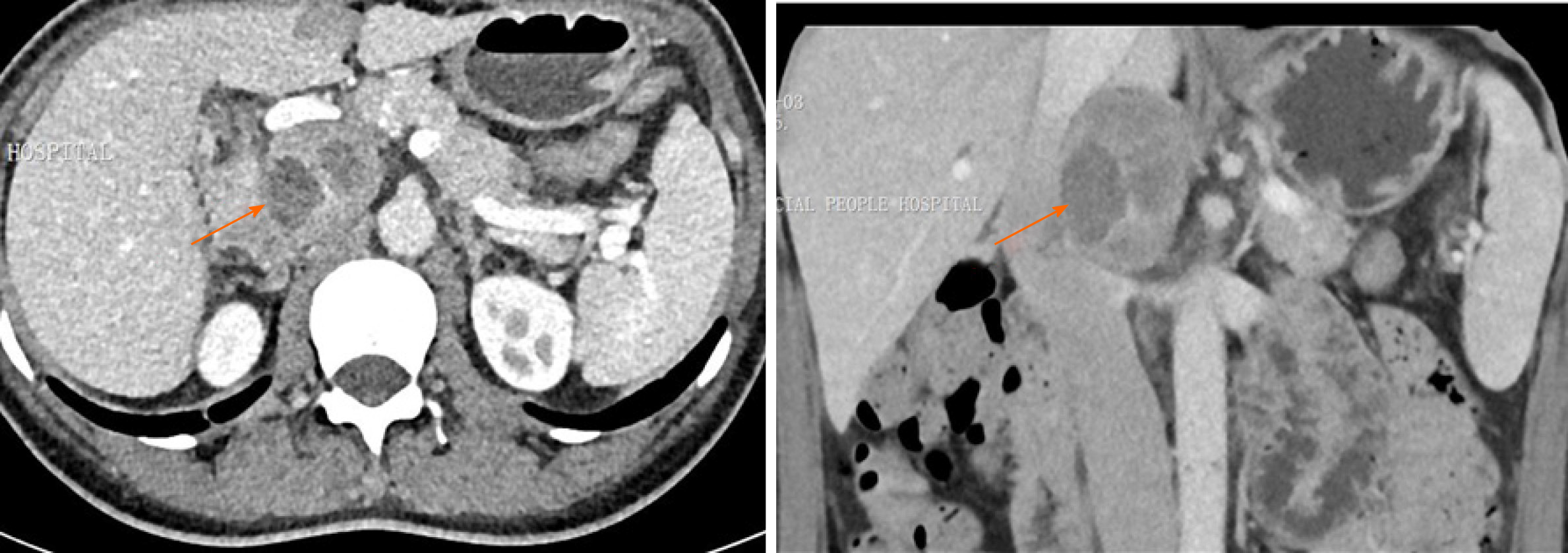
Figure 3 Contrast-enhanced computed tomography demonstrated an inhomogeneous right adrenal mass (6.
1 cm × 3.9 cm, orange arrows).
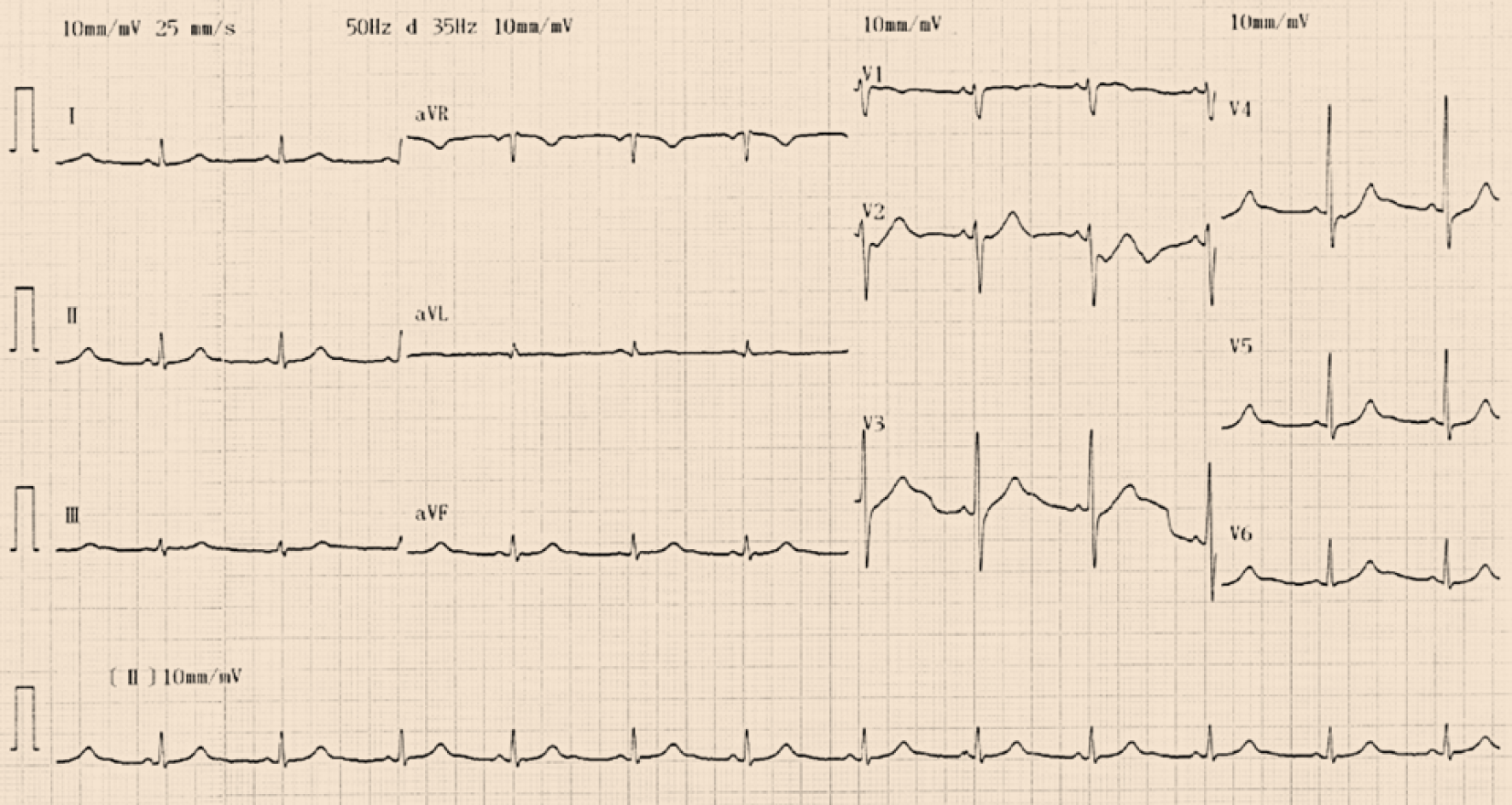
Figure 4 Twelve-lead electrocardiogram indicated QT prolongation and ST-segment depression in leads II, III, aVF and V3-V6 disappeared at the 1-mo follow-up after pheochromocytoma resection.
- Citation: Wu HY, Cao YW, Gao TJ, Fu JL, Liang L. Pheochromocytoma in a 49-year-old woman presenting with acute myocardial infarction: A case report. World J Clin Cases 2021; 9(15): 3752-3757
- URL: https://www.wjgnet.com/2307-8960/full/v9/i15/3752.htm
- DOI: https://dx.doi.org/10.12998/wjcc.v9.i15.3752


