Copyright
©The Author(s) 2021.
World J Clin Cases. May 26, 2021; 9(15): 3689-3695
Published online May 26, 2021. doi: 10.12998/wjcc.v9.i15.3689
Published online May 26, 2021. doi: 10.12998/wjcc.v9.i15.3689
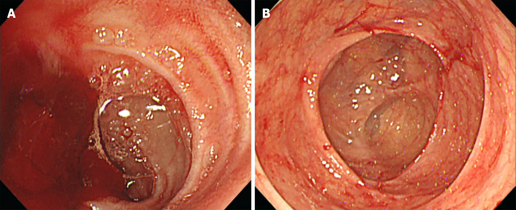
Figure 1 Initial endoscopic findings.
A: Lower gastrointestinal endoscopic findings show the possibility of small bowel bleeding; B: No evidence of bleeding was observed in the ascending colon and cecum.
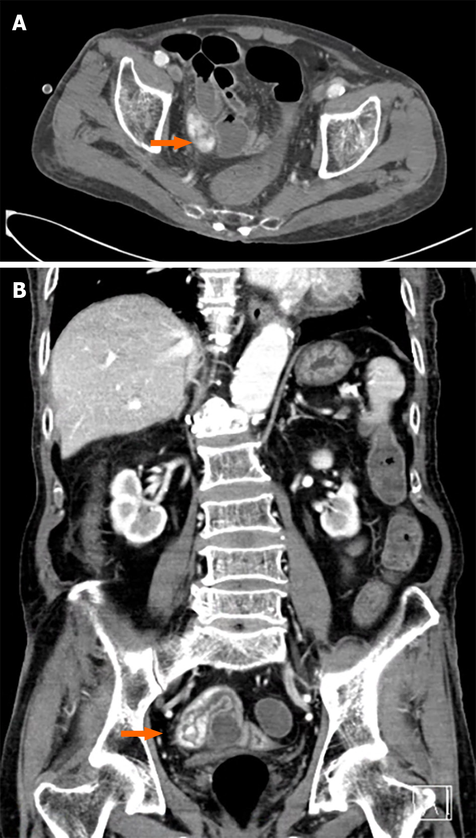
Figure 2 Alternate prism cover test findings.
A: Extravasation of contrast media (orange arrow) in the ileum cross-section view; B: Sagittal view.
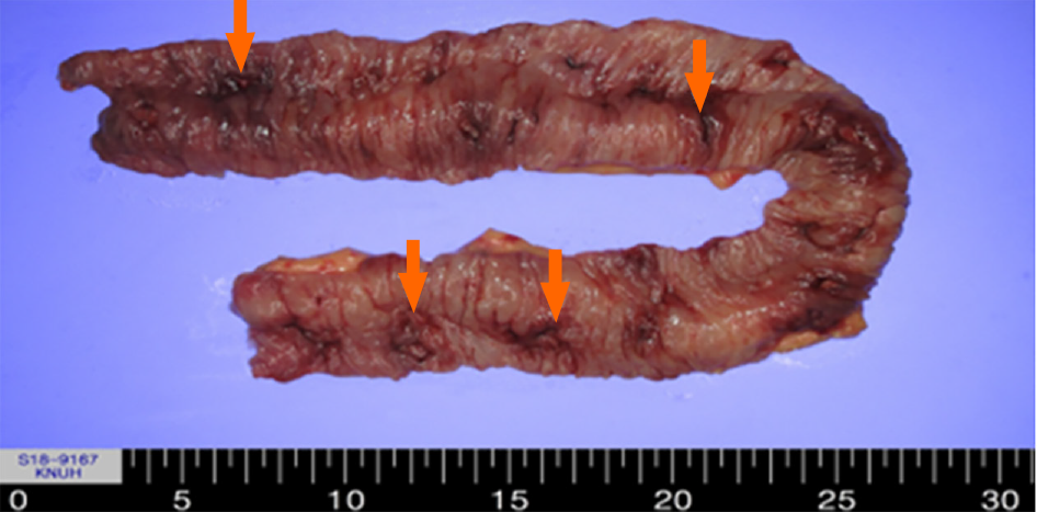
Figure 3 Excised specimen of the ileum, measuring 50 cm in length with multiple ulcerative lesions (orange arrow).
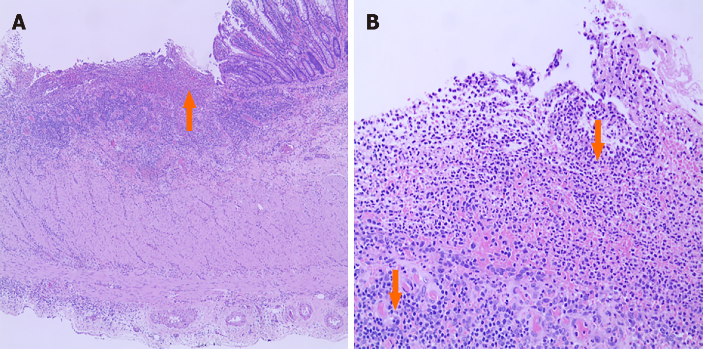
Figure 4 Histopathologic findings in the ileum.
A: Ulcer with inflammatory ulcer debris (orange arrow) [hematoxylin and eosin (H&E) stain, × 40]; B: New vascular proliferation consistent with granulation tissue formation (arrow) (H&E stain, × 200).
Figure 5 Capsule endoscopic findings one year after surgery.
The previous multiple ulcers were not observed.
- Citation: Lee SH, Ryu DR, Lee SJ, Park SC, Cho BR, Lee SK, Choi SJ, Cho HS. Small bowel ulcer bleeding due to suspected clopidogrel use in a patient with clopidogrel resistance: A case report. World J Clin Cases 2021; 9(15): 3689-3695
- URL: https://www.wjgnet.com/2307-8960/full/v9/i15/3689.htm
- DOI: https://dx.doi.org/10.12998/wjcc.v9.i15.3689


