Copyright
©The Author(s) 2021.
World J Clin Cases. May 26, 2021; 9(15): 3668-3674
Published online May 26, 2021. doi: 10.12998/wjcc.v9.i15.3668
Published online May 26, 2021. doi: 10.12998/wjcc.v9.i15.3668
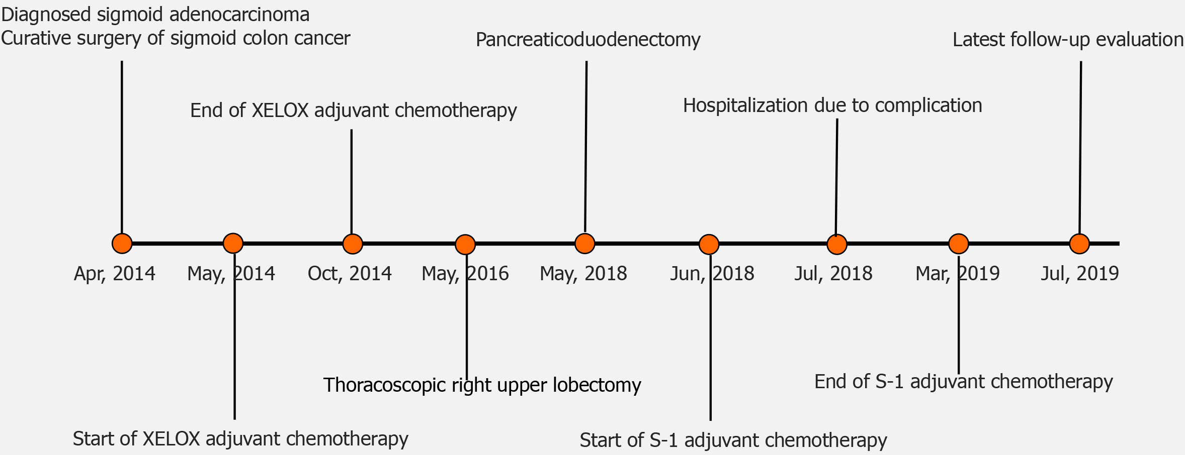
Figure 1 Course of disease management for the case described in this report.
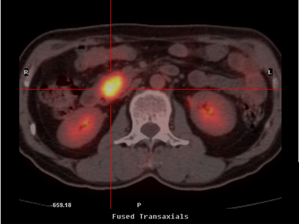
Figure 2 Positron emission tomography/computed tomography scan showing pathological hypermetabolism in the head of the pancreas.
No abnormalities were noted in the stomach, duodenum, common bile duct, or main pancreatic duct.
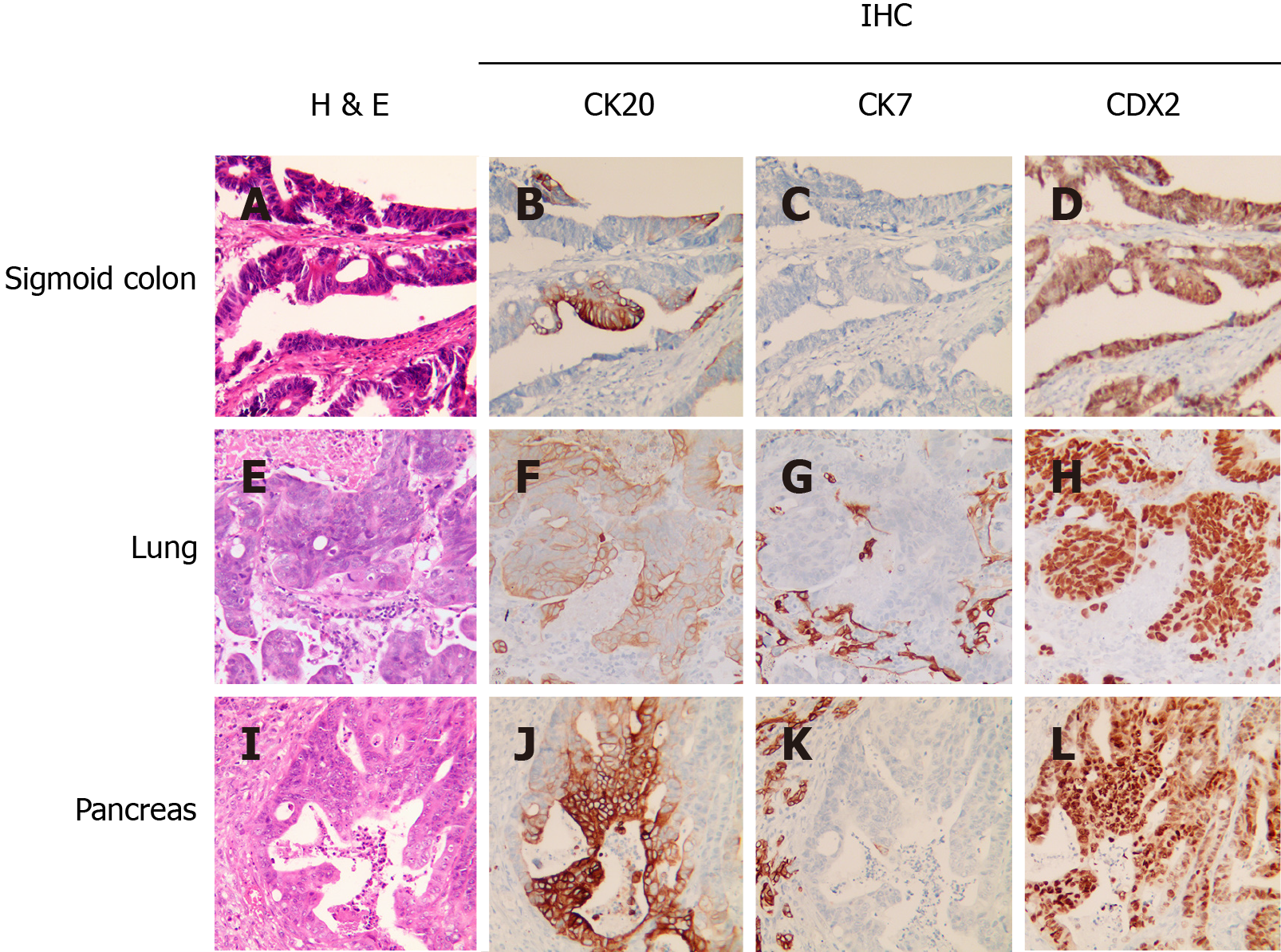
Figure 3 Representative results of hematoxylin and eosin and immunohistochemical staining of specimens from primary and metastatic lesions.
A-L: In the primary sigmoid adenocarcinoma, H&E staining (A) and CK20+ (B), CK7- (C), and CDX2+ (D) with IHC were similar to findings in lung metastasis (E-H) of CK20+ (F), CK7- (G), and CDX2+ (H). In addition, the pancreatic metastasis (I-L) also displayed biomarker expression profile consistent with colon cancer as the tissue of origin. H&E: Hematoxylin and eosin; IHC: Immunohistochemical.
- Citation: Yang J, Tang YC, Yin N, Liu W, Cao ZF, Li X, Zou X, Zhang ZX, Zhou J. Metachronous pulmonary and pancreatic metastases arising from sigmoid colon cancer: A case report. World J Clin Cases 2021; 9(15): 3668-3674
- URL: https://www.wjgnet.com/2307-8960/full/v9/i15/3668.htm
- DOI: https://dx.doi.org/10.12998/wjcc.v9.i15.3668


