Copyright
©The Author(s) 2021.
World J Clin Cases. May 6, 2021; 9(13): 3219-3226
Published online May 6, 2021. doi: 10.12998/wjcc.v9.i13.3219
Published online May 6, 2021. doi: 10.12998/wjcc.v9.i13.3219
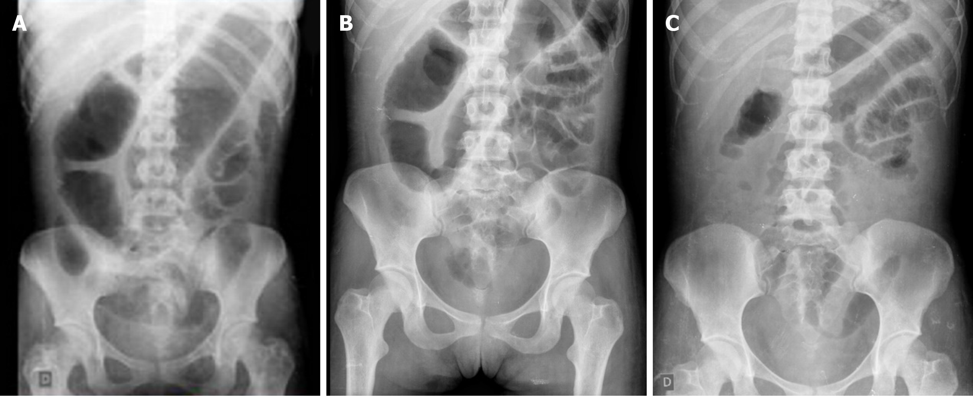
Figure 1 Abdominal X-ray during hospitalization.
A: Abdominal X-ray at admission (day 1) evidenced colonic dilation of 7 cm, consistent with the diagnosis of megacolon; B: Abdominal X-ray during treatment with oral vancomycin and hydrocortisone (day 4); C: Abdominal X-ray on the third day of treatment with hydrocortisone evidencing improvement of colonic distention (day 6).
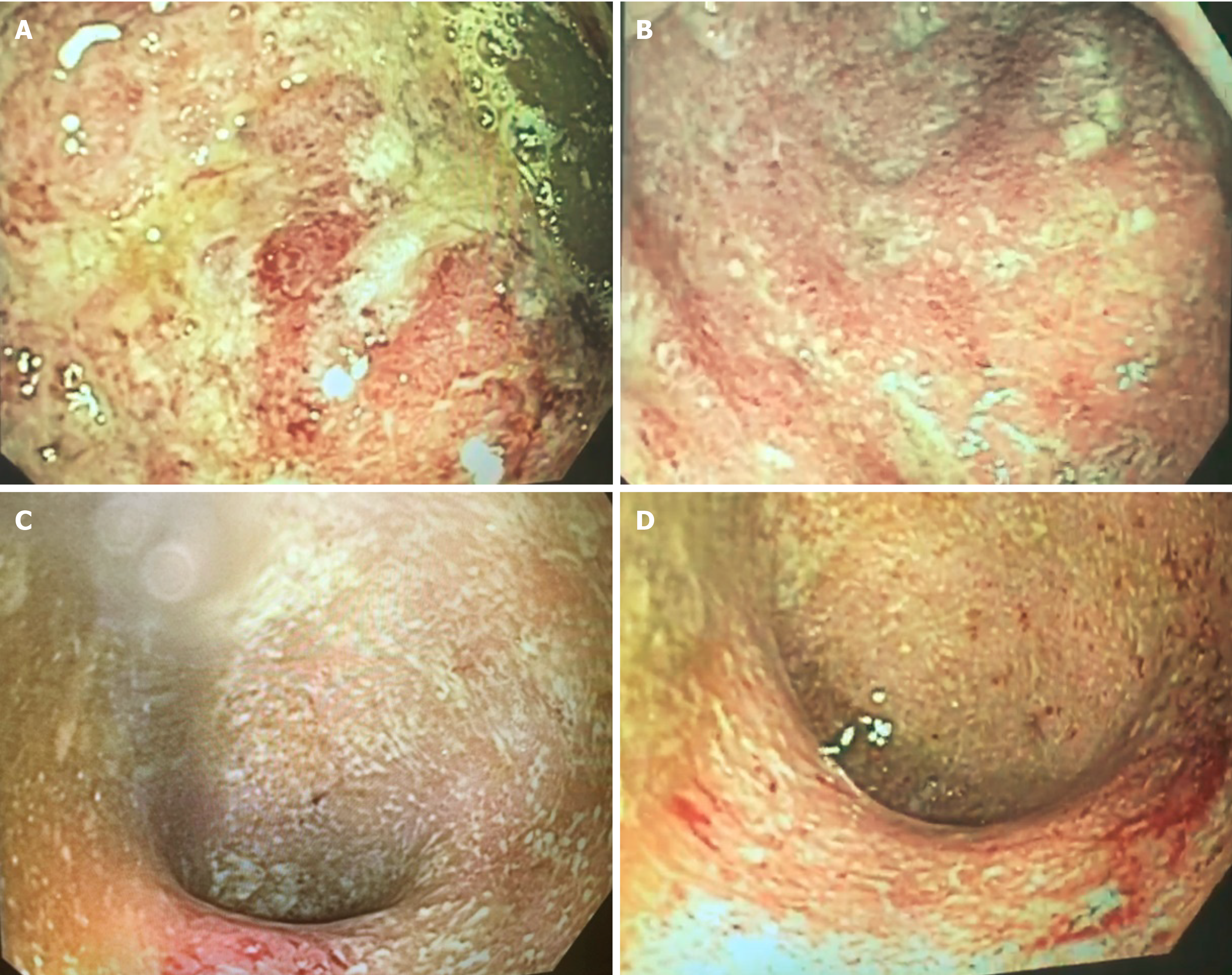
Figure 2 Flexible sigmoidoscopy performed during hospitalization.
A-D: Endoscopic image showing ulcers covered by fibrin, friability, edema and marked enanthem with spontaneous bleeding in sigmoid (A and B) and rectum (C and D), consistent with the ulcerative colitis of severe activity (Mayo endoscopic score 3).
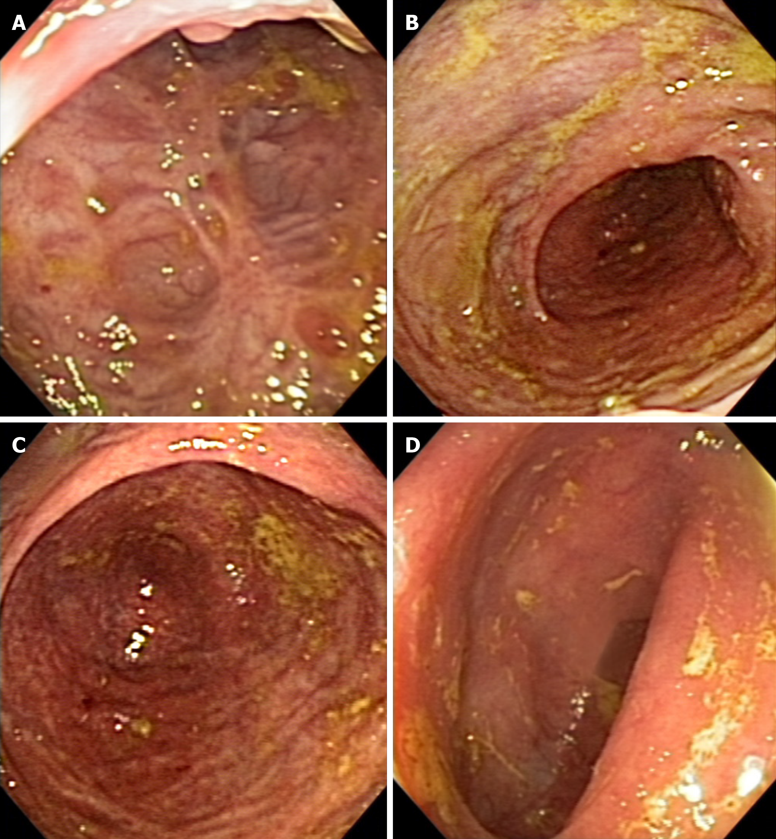
Figure 3 Colonoscopy performed 6 mo after starting the treatment with infliximab.
A-D: Endoscopic image showing discrete erythema and decreased vascular pattern, consistent with the ulcerative colitis of mild activity (Mayo endoscopic score 1).
- Citation: Garate ALSV, Rocha TB, Almeida LR, Quera R, Barros JR, Baima JP, Saad-Hossne R, Sassaki LY. Treatment of acute severe ulcerative colitis using accelerated infliximab regimen based on infliximab trough level: A case report. World J Clin Cases 2021; 9(13): 3219-3226
- URL: https://www.wjgnet.com/2307-8960/full/v9/i13/3219.htm
- DOI: https://dx.doi.org/10.12998/wjcc.v9.i13.3219


