Copyright
©The Author(s) 2021.
World J Clin Cases. May 6, 2021; 9(13): 3212-3218
Published online May 6, 2021. doi: 10.12998/wjcc.v9.i13.3212
Published online May 6, 2021. doi: 10.12998/wjcc.v9.i13.3212
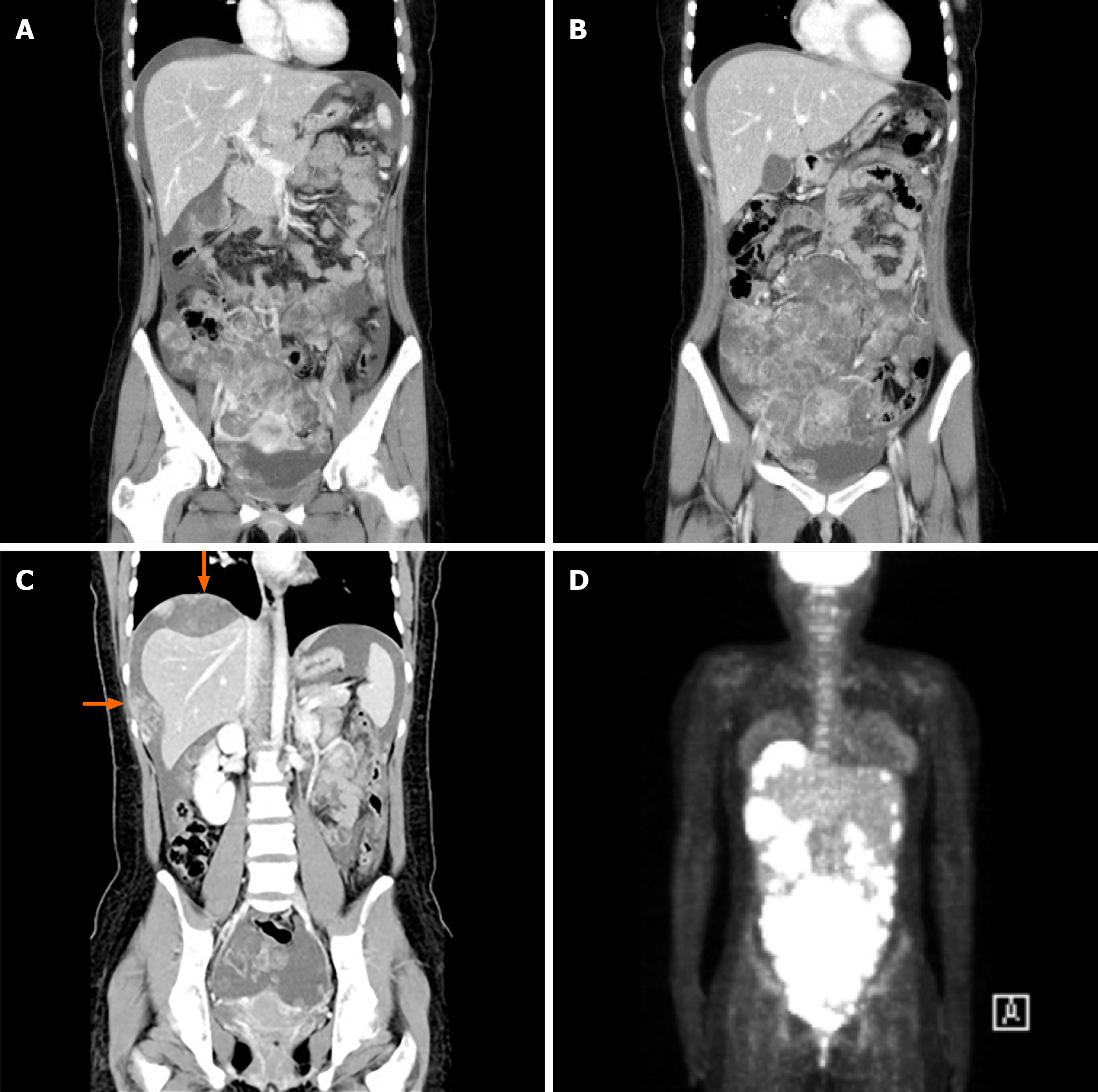
Figure 1 Imaging studies performed after hospitalization.
A-C: Abdominal contrast-enhanced computed tomography reveals a large amount of ascites with multifocal irregular arterial enhancing masses and nodules in the right adnexa with uterus (A), omentum and mesentery (B), and diaphragm and peritoneal wall in perihepatic space (arrows) (C); D: Positron emission tomography-computed tomography reveals multifocal hypermetabolism that spread extensively in the peritoneal cavity.
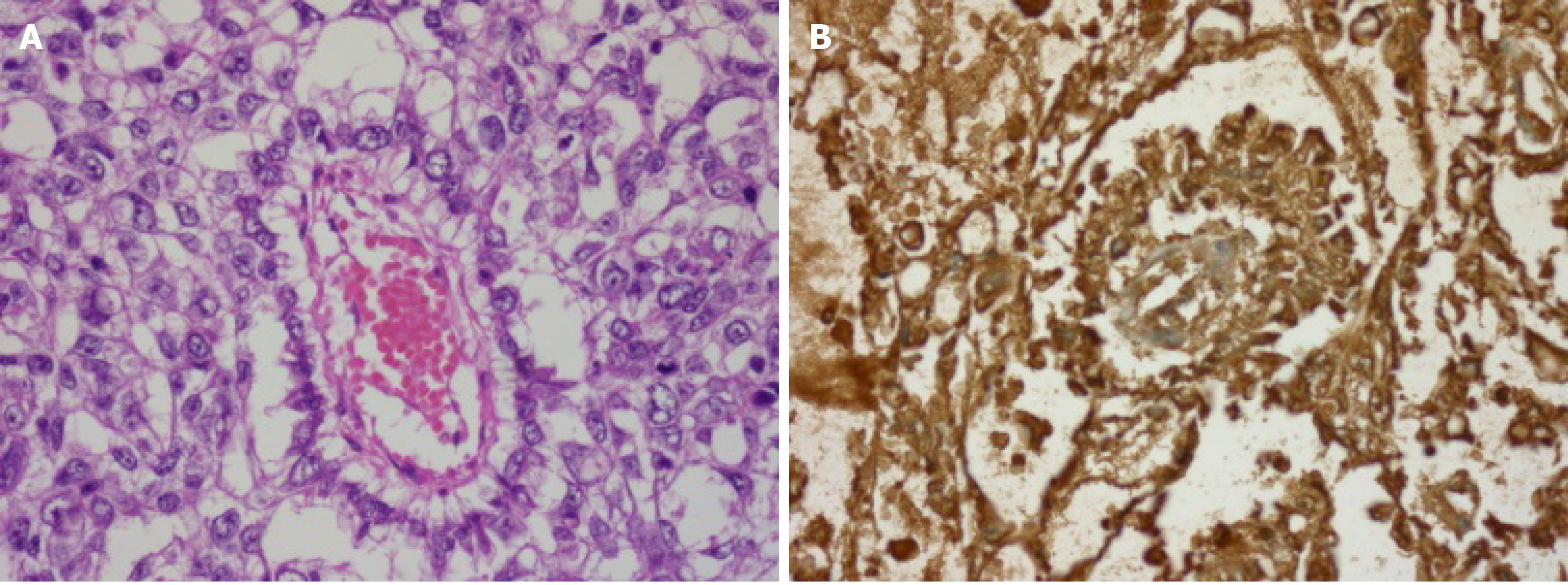
Figure 2 Pathology image.
A: Photomicrograph shows cuboidal-shaped tumor cells around central vessel forming the characteristic SchillerDuval body (hematoxylin and eosin, × 400); B: Immunohistochemical staining was positive for alpha-fetoprotein (original magnification × 400).
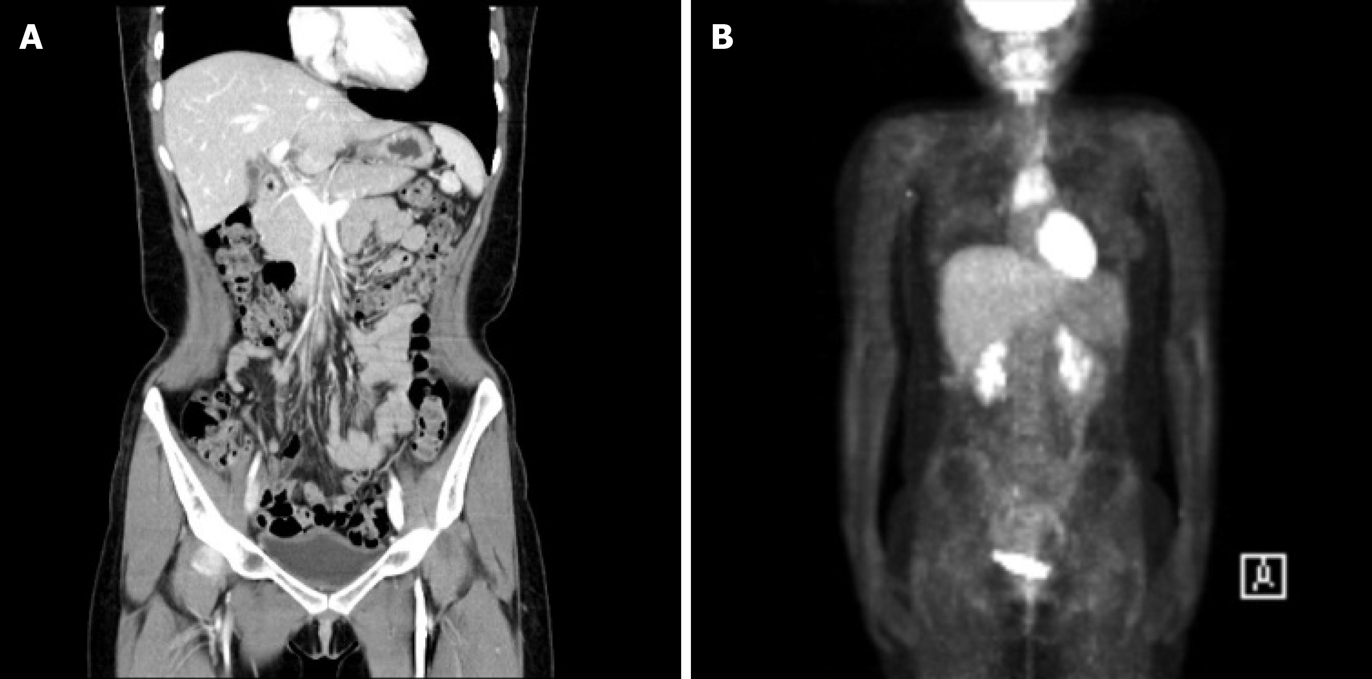
Figure 3 Imaging studies performed 6 year after treatment.
A: No definite evidence of local recurrence in abdominal contrast-enhanced computed tomography; B: No evidence of tumor recurrence nor distant metastasis in positron emission tomography-computed tomography.
- Citation: Oh HK, Park SN, Kim BR. Laparoscopic uncontained power morcellation-induced dissemination of ovarian endodermal sinus tumors: A case report. World J Clin Cases 2021; 9(13): 3212-3218
- URL: https://www.wjgnet.com/2307-8960/full/v9/i13/3212.htm
- DOI: https://dx.doi.org/10.12998/wjcc.v9.i13.3212


