Copyright
©The Author(s) 2021.
World J Clin Cases. May 6, 2021; 9(13): 3177-3184
Published online May 6, 2021. doi: 10.12998/wjcc.v9.i13.3177
Published online May 6, 2021. doi: 10.12998/wjcc.v9.i13.3177
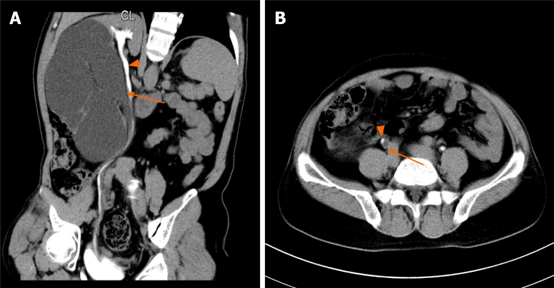
Figure 1 Duplicate renal malformation confirmed by computerized tomography urography.
A and B: The patient has a duplication of renal malformation on the right side, with independent ureters in the upper (orange arrows) and lower parts (orange triangle), and the lower kidney is a malformed kidney with hydrops.
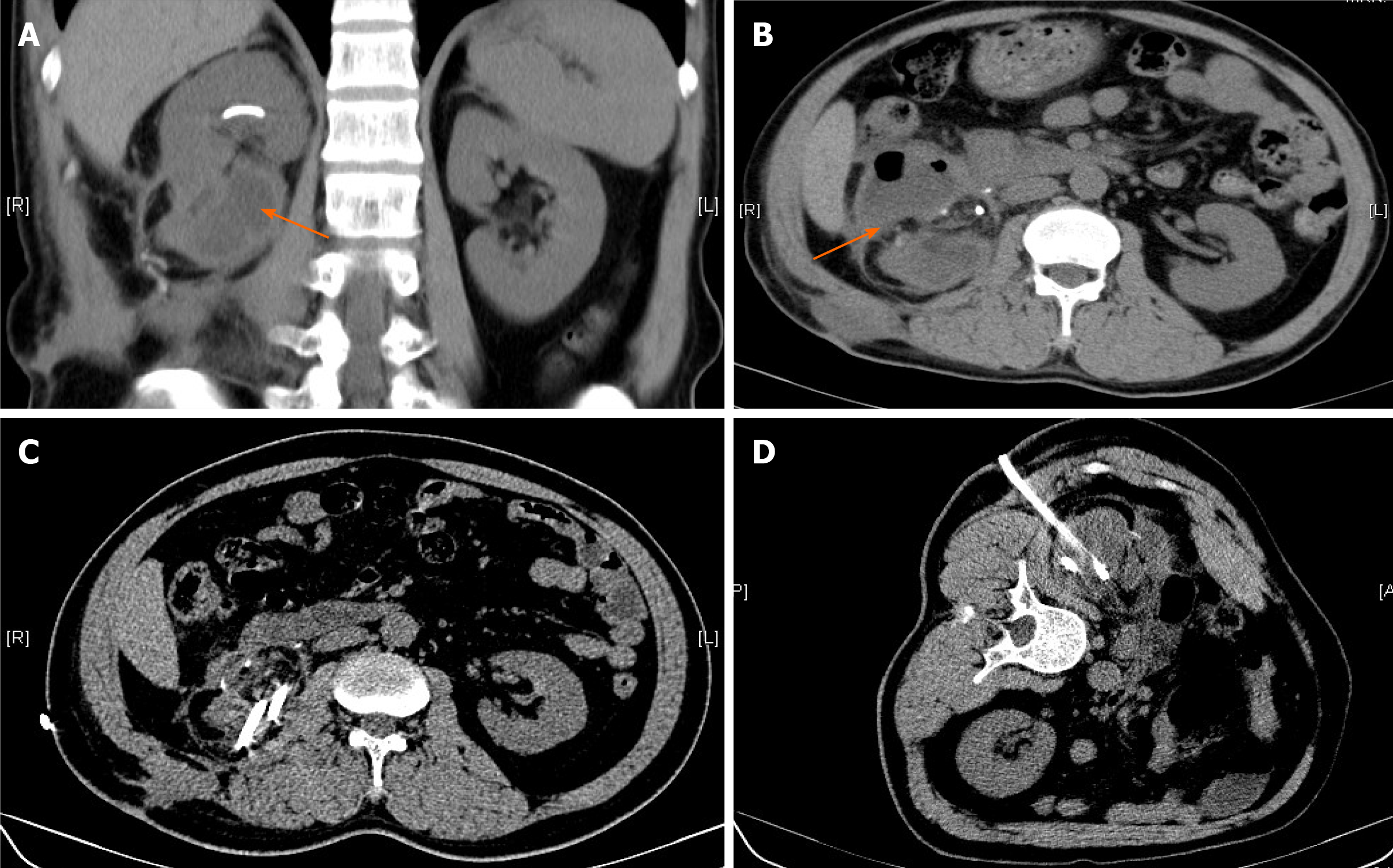
Figure 2 Perinephric effusion after heminephrectomy of computed tomography.
A and B: Shown is the perinephric effusion (orange arrows) in the lower pole of the right kidney after the operation; C and D: Shown is puncture drainage of right perinephric effusion.
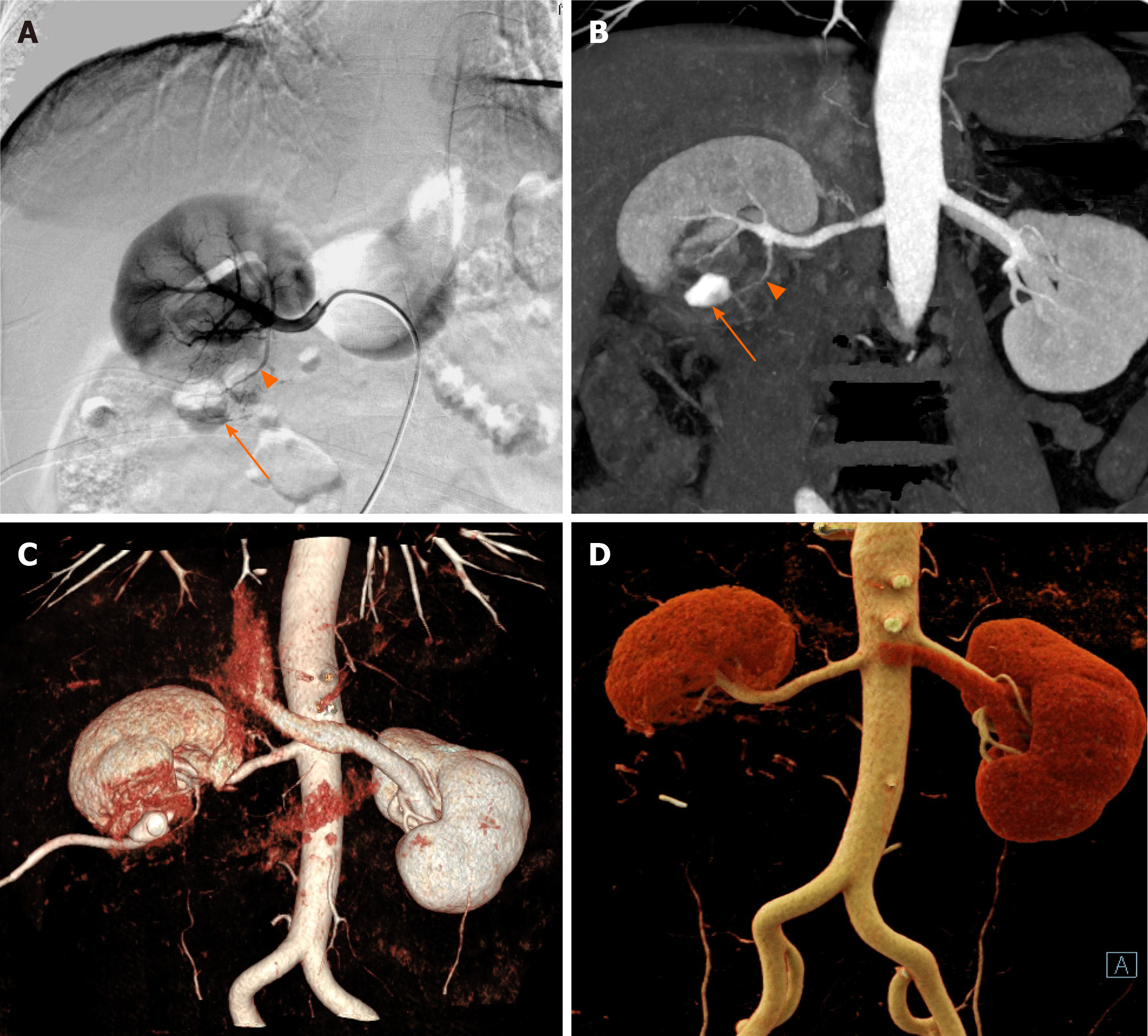
Figure 3 Residual kidney section confirmed by computed tomography angiography and digital subtraction angiography.
A and B: The patient's preoperative computed tomography angiography and digital subtraction angiography comparison confirmed that a branch artery (orange triangle) in the lower pole of the right kidney runs out of the contour of the kidney to supply blood to the residual kidney section (orange arrows); C and D: The patient’s preoperative and postoperative computed tomography angiography comparisons confirmed that only the main trunk remained after interventional embolization of the residual renal artery branch.
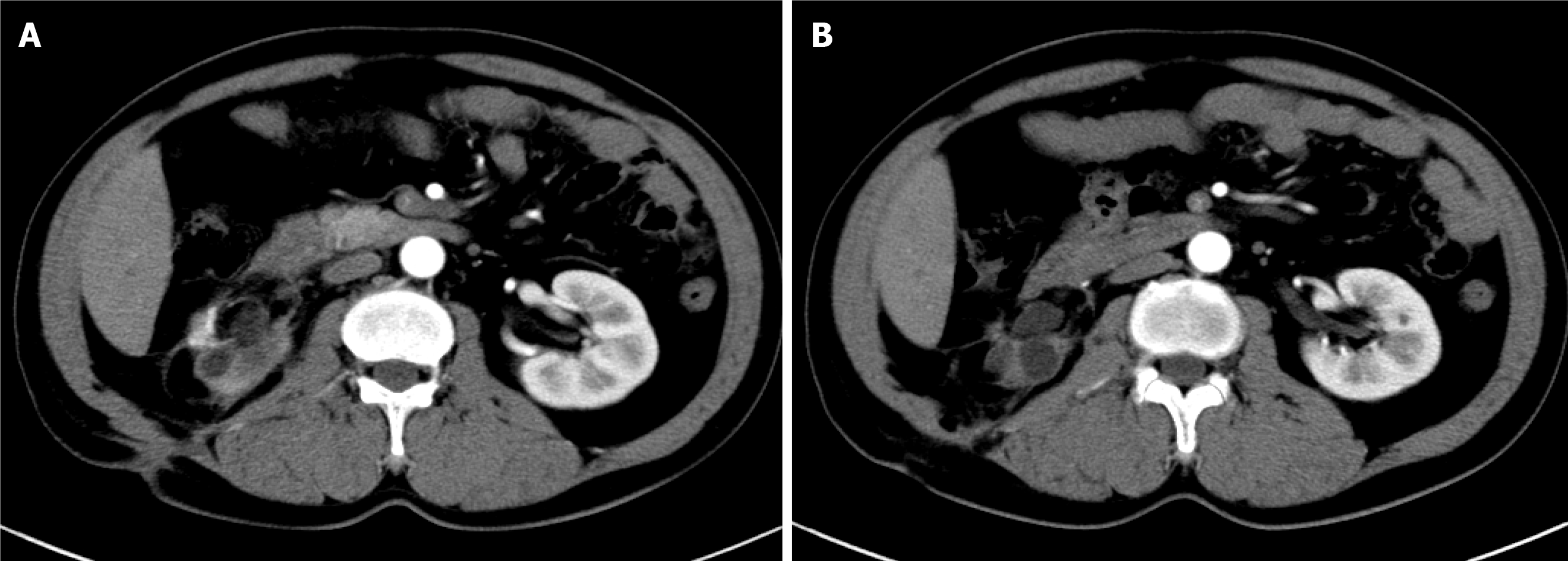
Figure 4 Abdominal computed tomography angiography 3 yr after surgery.
A and B: The patient was admitted to the hospital in January 2018, and the computed tomography showed that the right perinephric effusion had disappeared.
- Citation: Yang T, Wen J, Xu TT, Cui WJ, Xu J. Renal artery embolization in the treatment of urinary fistula after renal duplication: A case report and review of literature. World J Clin Cases 2021; 9(13): 3177-3184
- URL: https://www.wjgnet.com/2307-8960/full/v9/i13/3177.htm
- DOI: https://dx.doi.org/10.12998/wjcc.v9.i13.3177


