Copyright
©The Author(s) 2021.
World J Clin Cases. May 6, 2021; 9(13): 3170-3176
Published online May 6, 2021. doi: 10.12998/wjcc.v9.i13.3170
Published online May 6, 2021. doi: 10.12998/wjcc.v9.i13.3170
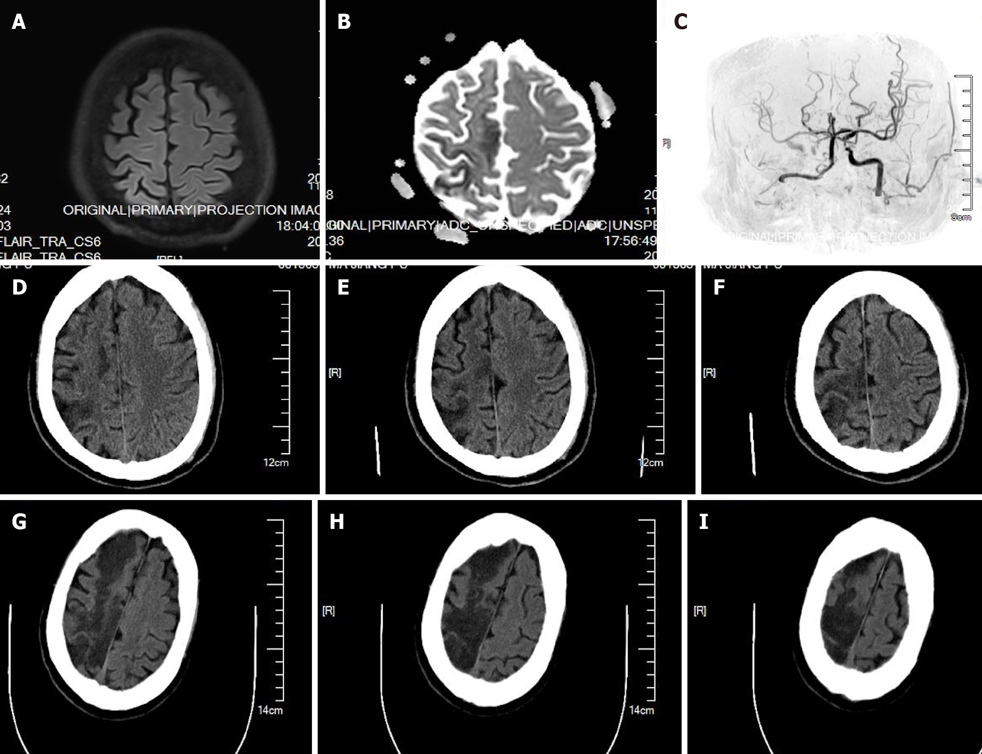
Figure 1 Magnetic resonance and computed tomography images of case 1.
A-C: Magnetic resonance imaging (A and B) and angiography (C) performed 4 h after surgery showed right cerebral infarction, occlusion of right internal carotid artery (ICA), and severe stenosis of left ICA; D-F: Computed tomography (CT) scans performed 1 d after surgery showed right cerebral infarction; G-I: CT scans performed 4 d after surgery showed right cerebral infarction progression.
- Citation: Jian MY, Liang F, Liu HY, Han RQ. Perioperative massive cerebral stroke in thoracic patients: Report of three cases. World J Clin Cases 2021; 9(13): 3170-3176
- URL: https://www.wjgnet.com/2307-8960/full/v9/i13/3170.htm
- DOI: https://dx.doi.org/10.12998/wjcc.v9.i13.3170


