Copyright
©The Author(s) 2021.
World J Clin Cases. May 6, 2021; 9(13): 3157-3162
Published online May 6, 2021. doi: 10.12998/wjcc.v9.i13.3157
Published online May 6, 2021. doi: 10.12998/wjcc.v9.i13.3157
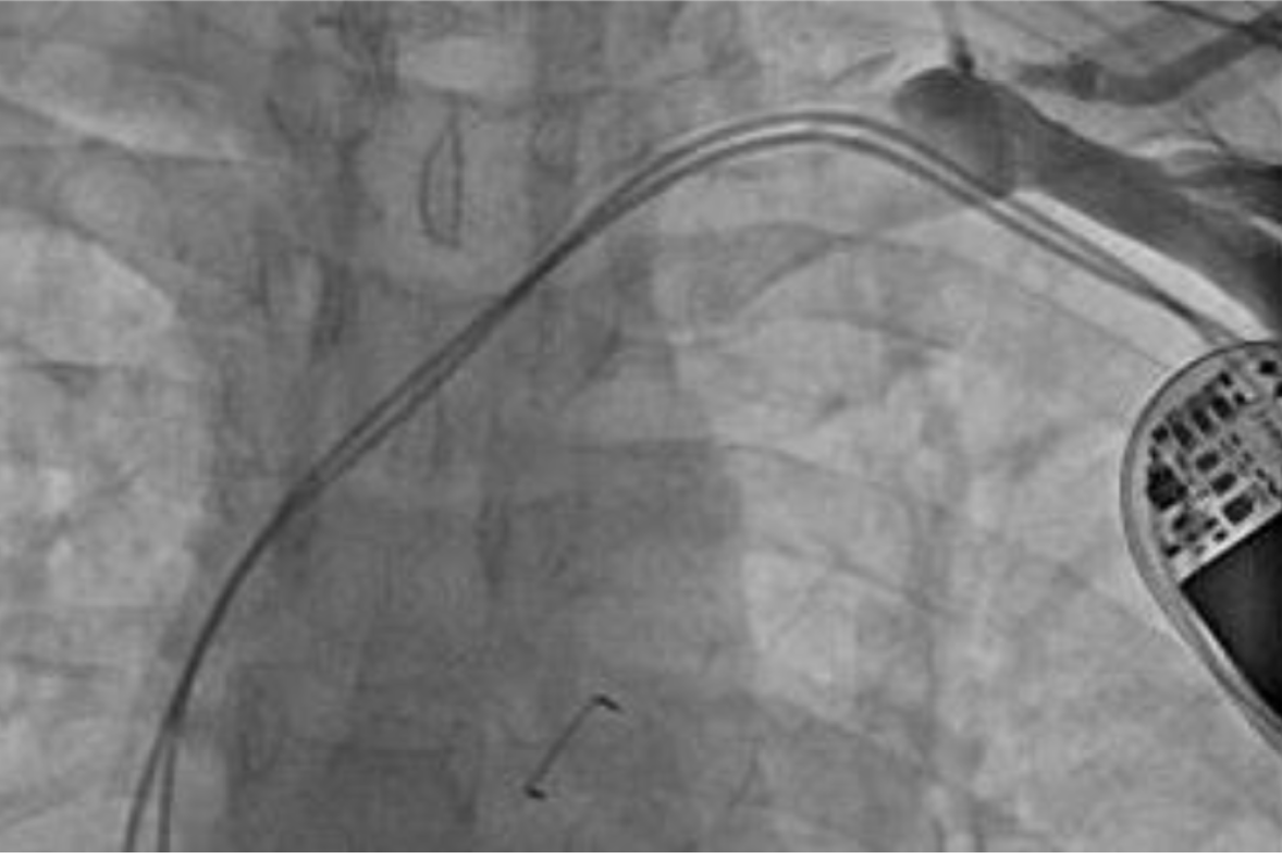
Figure 1 Venogram showing complete occlusion of the left subclavian vein.
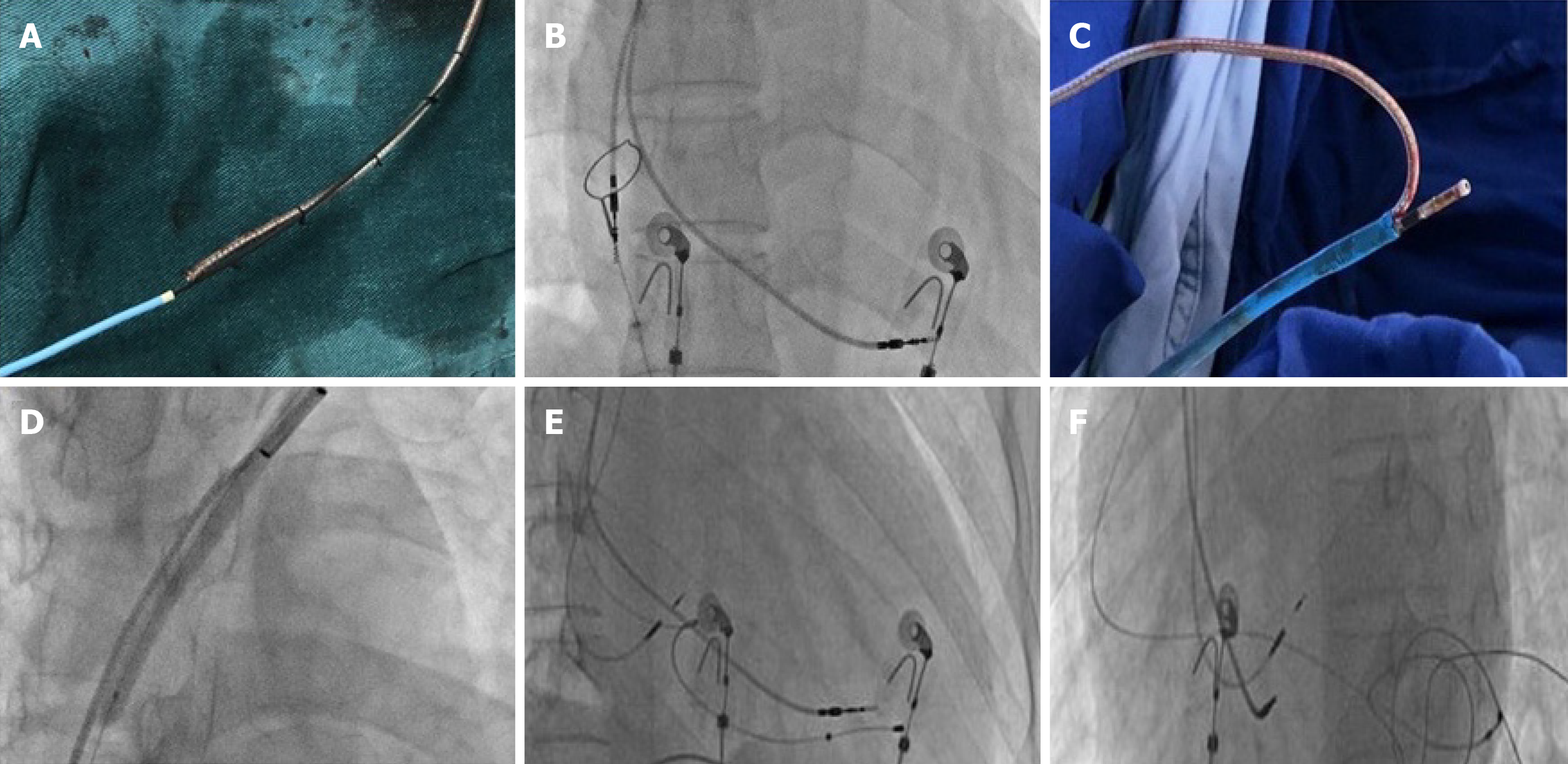
Figure 2 Cardiac resynchronization therapy pacemaker procedure.
A: Proximally cut lead portions and a Radifocus guidewire tip were fixed with a suture, and the rest of the wire was covered by a Finecross catheter; B: The atrial lead tip captured in a goose-neck snare was positioned in the right atrium; C: The atrial lead was placed in the 8 French femoral vein sheath and removed from the right femoral vein; D: The balloon was dilated across the occlusion in the left subclavian vein; E and F: Intra-procedure fluoroscopy showed the location of the lead of the cardiac resynchronization therapy pacemaker.
- Citation: Zhong JY, Zheng XW, Li HD, Jiang LF. Successful upgrade to cardiac resynchronization therapy for cardiac implantation-associated left subclavian vein occlusion: A case report. World J Clin Cases 2021; 9(13): 3157-3162
- URL: https://www.wjgnet.com/2307-8960/full/v9/i13/3157.htm
- DOI: https://dx.doi.org/10.12998/wjcc.v9.i13.3157


