Copyright
©The Author(s) 2021.
World J Clin Cases. May 6, 2021; 9(13): 3095-3101
Published online May 6, 2021. doi: 10.12998/wjcc.v9.i13.3095
Published online May 6, 2021. doi: 10.12998/wjcc.v9.i13.3095
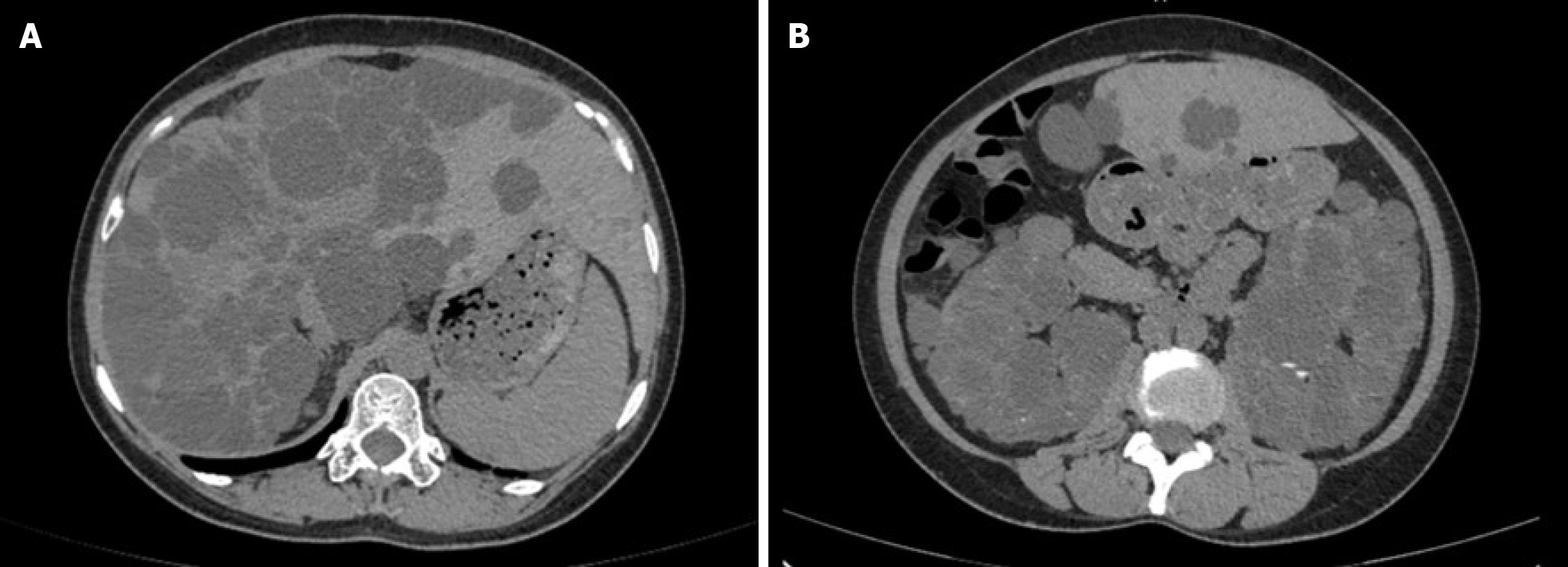
Figure 1 Computed tomography showed the imaging manifestations of polycystic liver and polycystic kidney.
A: Polycystic liver; B: Polycystic kidney.
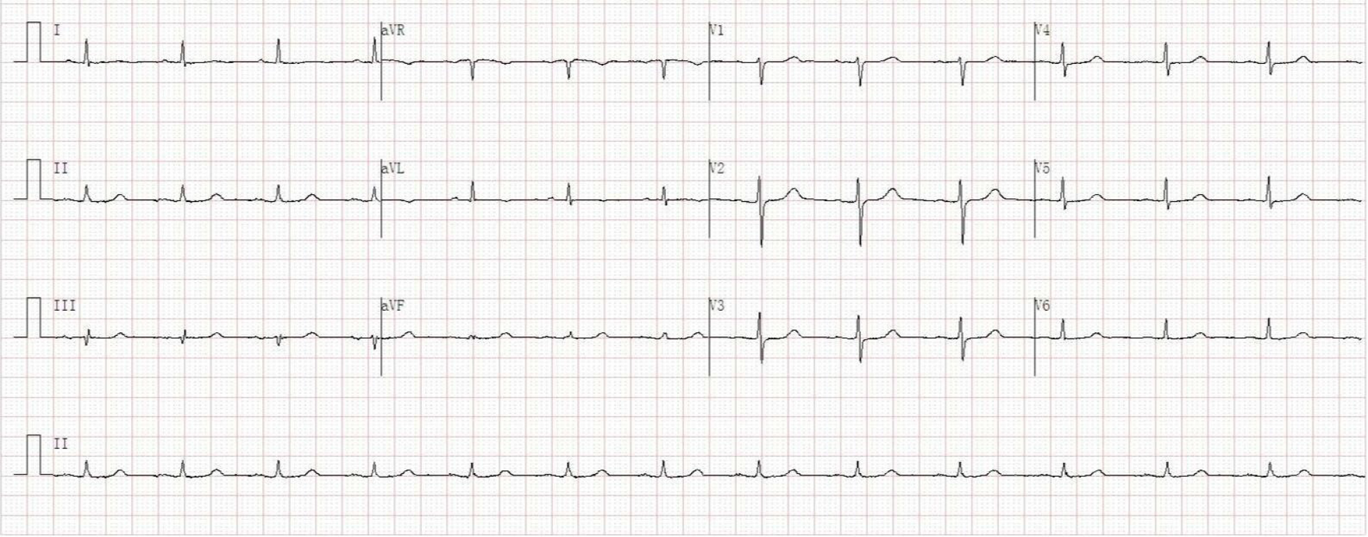
Figure 2
Initial electrocardiogram in the emergency room.
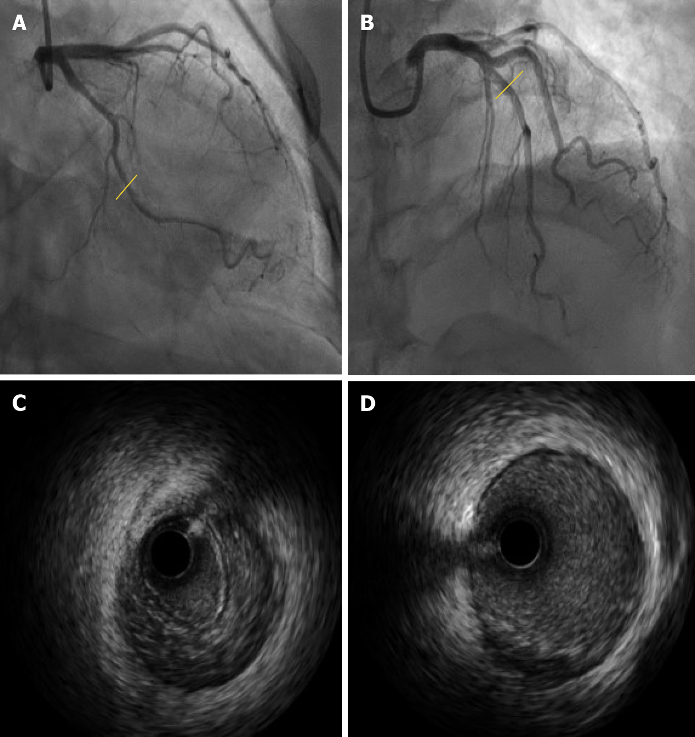
Figure 3 Coronary angiography and intravascular ultrasound images.
A and B: Coronary angiography revealed no clear signs of dissection in the left circumflex branch (A) and left anterior descending branch (B); C and D: Intravascular ultrasound showed clear signs of dissection (intramural hematoma: C) of the left circumflex branch, and atherosclerotic plaque formation in the left anterior descending branch (D).
- Citation: Qian J, Lai Y, Kuang LJ, Chen F, Liu XB. Spontaneous coronary dissection should not be ignored in patients with chest pain in autosomal dominant polycystic kidney disease: A case report. World J Clin Cases 2021; 9(13): 3095-3101
- URL: https://www.wjgnet.com/2307-8960/full/v9/i13/3095.htm
- DOI: https://dx.doi.org/10.12998/wjcc.v9.i13.3095


