Copyright
©The Author(s) 2021.
World J Clin Cases. May 6, 2021; 9(13): 3063-3069
Published online May 6, 2021. doi: 10.12998/wjcc.v9.i13.3063
Published online May 6, 2021. doi: 10.12998/wjcc.v9.i13.3063
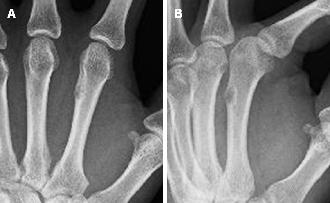
Figure 1 Radiographs obtained from a 40-year-old man who presented with pain in the left hand.
A: Antero-posterior view; B: lateral view radiographs revealed intracortical lucency and cortical thickening of the second metacarpal bone were observed on plain radiographs.
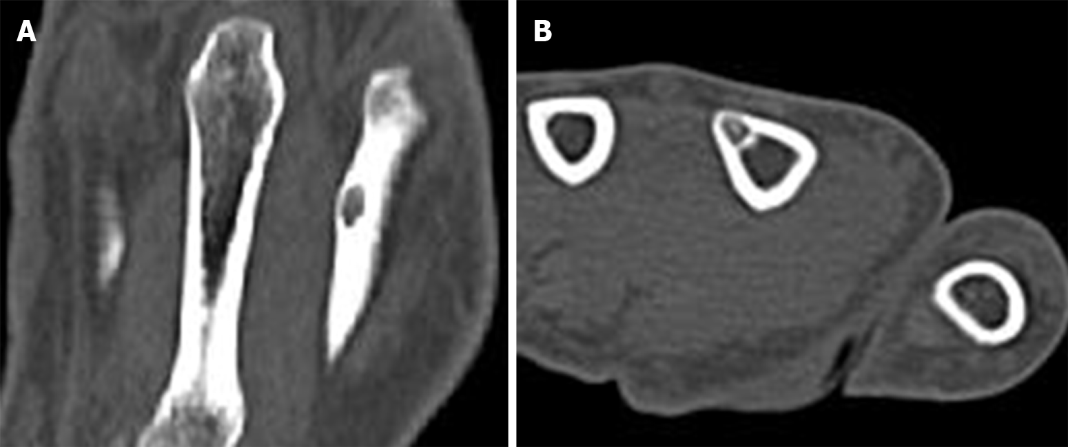
Figure 2 Plain computed tomography images.
A: Coronal view; B: Axial view images revealed a 4 mm, well-defined lytic lesion and intra-tumoral punctate calcification. The appearance of this lesion on computed tomography images is almost similar to that of osteoid osteoma.
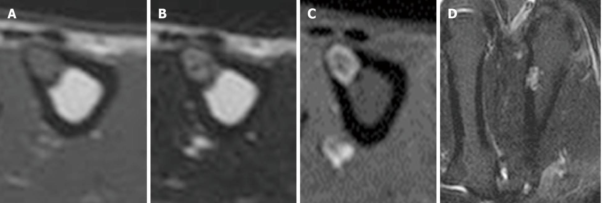
Figure 3 Magnetic resonance imaging of the second metacarpal bone.
A: Low signal intensity on axial T1-weighted images; B: High signal intensity on axial T2-weighted images; C: Axial contrast-enhanced magnetic resonance imaging. Only the tumor margin was enhanced; D: Coronal short tau inversion recovery magnetic resonance imaging.
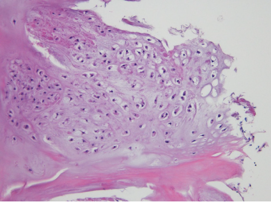
Figure 4 Histopathological image of the tumor.
Hypocellular, cytologically banal hyaline cartilage suggested chondroma and was located in the cortical bone. The sample was stained with hematoxylin-eosin and imaged at × 100 magnification.
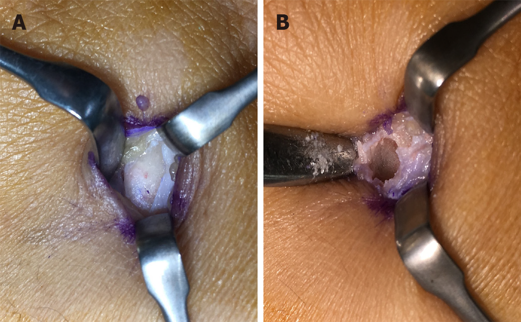
Figure 5 Photograph of the intraoperative appearance of the second metacarpal bone.
A: Before curettage, protrusion of cortical bone is revealed; B: After curettage, the lesion was completely embedded in the cortical bone.
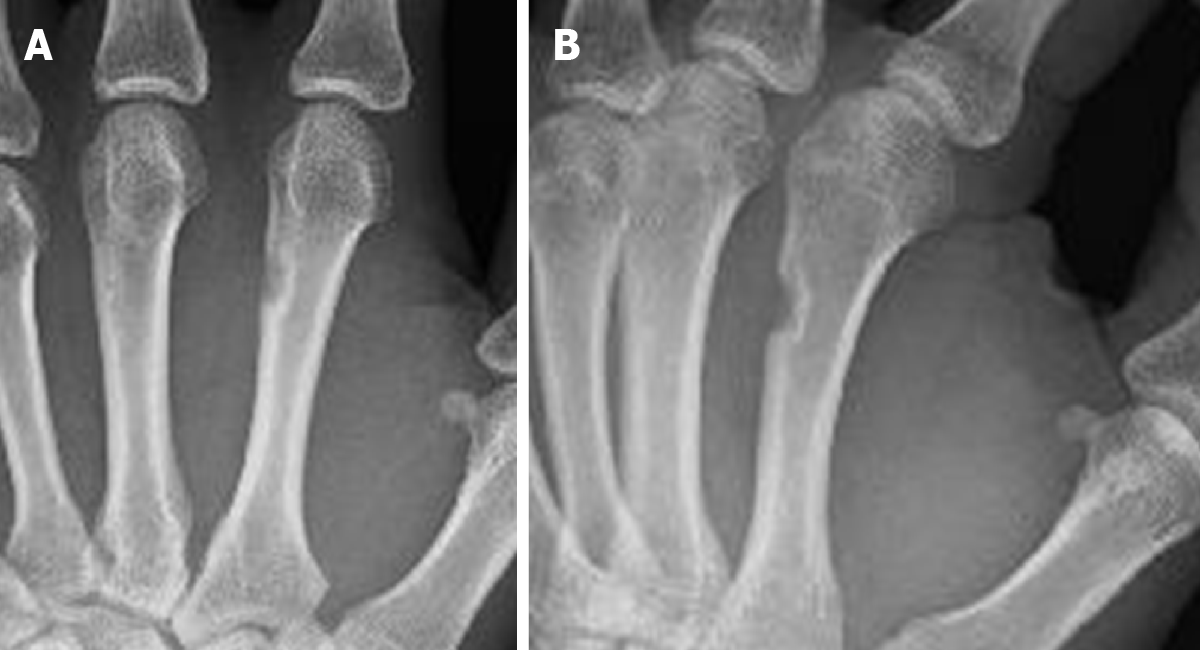
Figure 6 Postoperative plain radiographs obtained 1 year after treatment with no recurrence.
A: Antero-posterior view; B: Lateral view.
- Citation: Yoshida Y, Anazawa U, Watanabe I, Hotta H, Aoyama R, Suzuki S, Nagura T. Intracortical chondroma of the metacarpal bone: A case report. World J Clin Cases 2021; 9(13): 3063-3069
- URL: https://www.wjgnet.com/2307-8960/full/v9/i13/3063.htm
- DOI: https://dx.doi.org/10.12998/wjcc.v9.i13.3063


