Copyright
©The Author(s) 2021.
World J Clin Cases. Apr 16, 2021; 9(11): 2655-2661
Published online Apr 16, 2021. doi: 10.12998/wjcc.v9.i11.2655
Published online Apr 16, 2021. doi: 10.12998/wjcc.v9.i11.2655
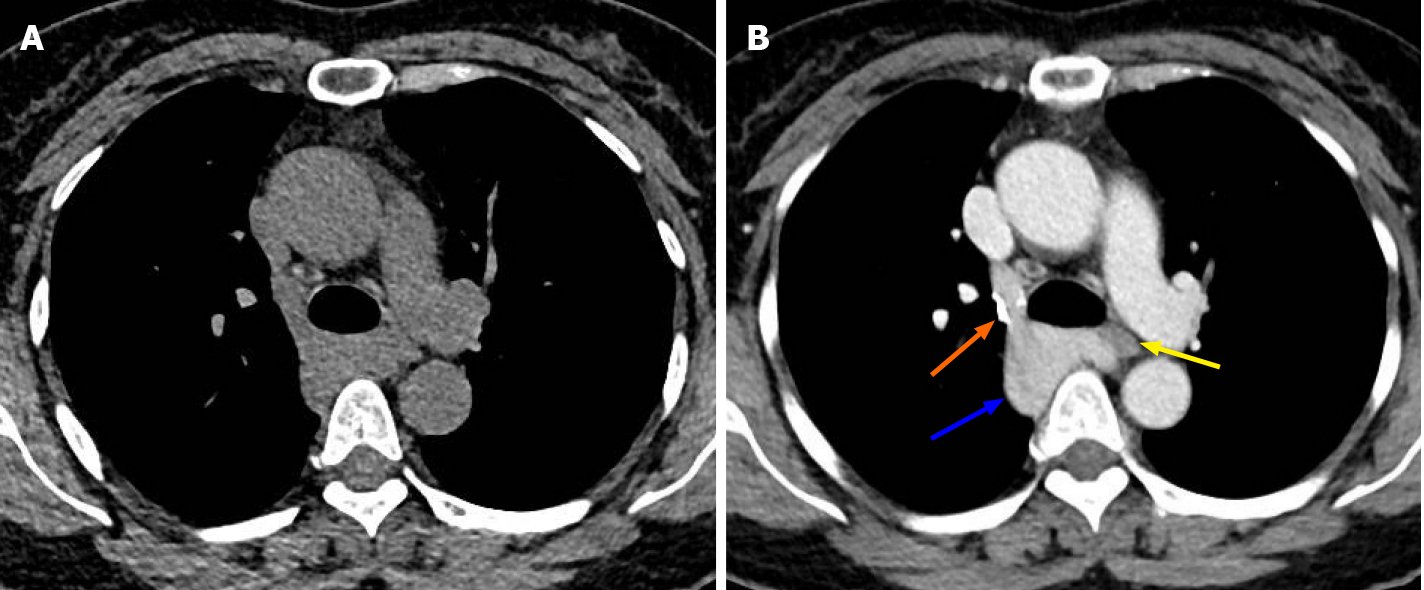
Figure 1 Tumor imaging by chest computed tomography.
A: Chest computed tomography showed a soft-tissue mass in the right posterior mediastinum; the mass was approximately 4.2 cm × 3.7 cm × 2.6 cm in size and showed unclear boundaries with the esophagus and compression of the trachea; B: Enhanced scanning showed that the azygos vein arch widened, the contrast agent remained in the azygos vein arch, the tumor exhibited delayed enhancement, and the internal density was uneven (blue arrow). In the venous phase, the tumor was connected to the superior vena cava, and the degree of enhancement was the same as that of the superior vena cava. The boundary between the tumor and the esophagus was clear. Note the azygos vein valve (orange arrow). The esophagus was compressed by the azygos vein aneurysm (yellow arrow).
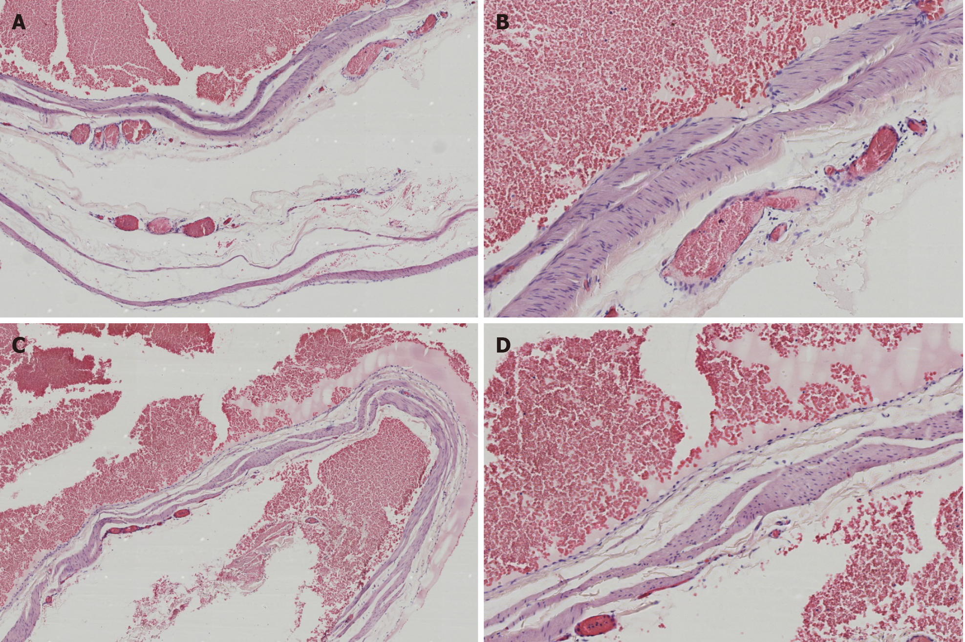
Figure 2 Hematoxylin and eosin staining.
A and B: There were enlarged and dilated thin-walled blood vessels with intact endothelial cells on the wall, in which the medial muscle layer was thinned, and a large number of red blood cells were found outside the lumen (A: 40 ×; B: 100 ×); C and D: Multiple red blood cells were observed in the expanded blood vessel wall, composing the mural thrombus (C: 40 ×; D: 100 ×).
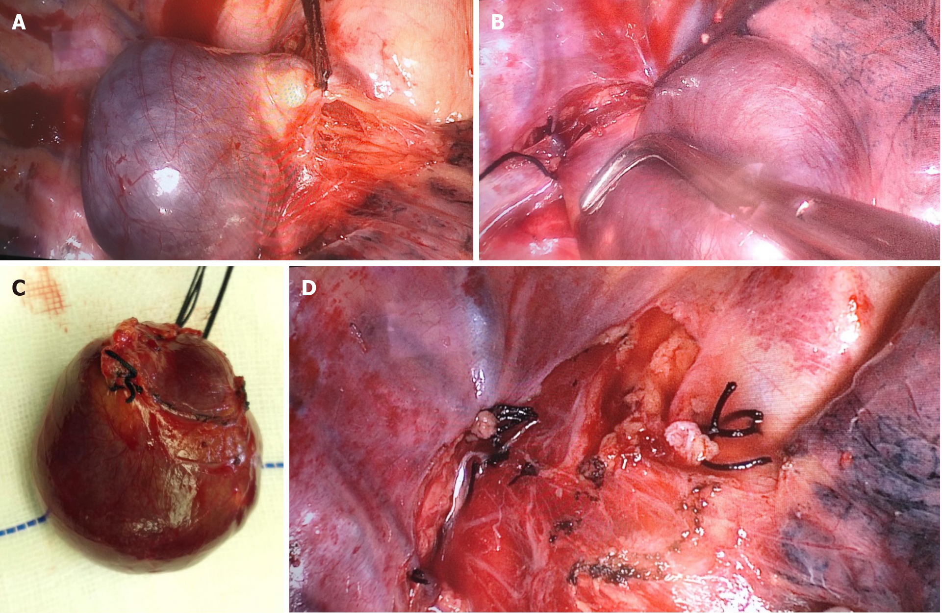
Figure 3 Intraoperative management.
A: The azygos vein arch was enlarged and showed dark purple cystic changes. The tumor was slightly adherent to the surrounding tissues. The azygos vein was ligated at the connection of the superior vena cava with silk thread; B: The distal vessel was ligated; C: A complete resection of the azygos vein aneurysm was performed; D: Shown is the intraoperative wound of the right posterior mediastinum.
- Citation: Wang ZX, Yang LL, Xu ZN, Lv PY, Wang Y. Surgical therapy for hemangioma of the azygos vein arch under thoracoscopy: A case report. World J Clin Cases 2021; 9(11): 2655-2661
- URL: https://www.wjgnet.com/2307-8960/full/v9/i11/2655.htm
- DOI: https://dx.doi.org/10.12998/wjcc.v9.i11.2655


