Copyright
©The Author(s) 2021.
World J Clin Cases. Apr 16, 2021; 9(11): 2641-2648
Published online Apr 16, 2021. doi: 10.12998/wjcc.v9.i11.2641
Published online Apr 16, 2021. doi: 10.12998/wjcc.v9.i11.2641
Figure 1 Tri-phasic, contrast-enhanced abdominal computer tomography.
There were heterogeneous masses in the liver with a maximal size of 8.3 cm at segment 4 (arrow). A: Contrast enhancement in the arterial phase; B: Contrast washout in the portal venous phase; C: Contrast washout in the delayed phase.
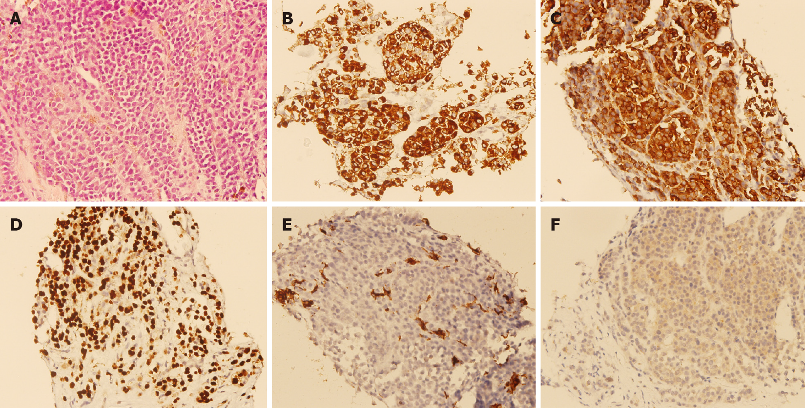
Figure 2 Pathological results of the lesions.
A: Pathological examinations showed epithelioid tumor cells with moderate nuclear atypia, and focal intracytoplasmic melanin pigments in solid nests infiltrating in the liver parenchyma (hematoxylin and eosin, 400 ×); B: Immunohistochemical staining revealed that the tumor cells were HMB45 positive [avidin-biotin complex (ABC), 400 ×]; C: Melan-A positive (ABC, 400 ×); D: SOX10 positive (ABC, 400 ×); E: cytokeratin negative (ABC, 400 ×); F: BRAF V600E positive (ABC, 400 ×).
- Citation: Cheng AC, Lin YJ, Chiu SH, Shih YL. Combined immune checkpoint inhibitors of CTLA4 and PD-1 for hepatic melanoma of unknown primary origin: A case report. World J Clin Cases 2021; 9(11): 2641-2648
- URL: https://www.wjgnet.com/2307-8960/full/v9/i11/2641.htm
- DOI: https://dx.doi.org/10.12998/wjcc.v9.i11.2641


