Copyright
©The Author(s) 2021.
World J Clin Cases. Apr 16, 2021; 9(11): 2619-2626
Published online Apr 16, 2021. doi: 10.12998/wjcc.v9.i11.2619
Published online Apr 16, 2021. doi: 10.12998/wjcc.v9.i11.2619
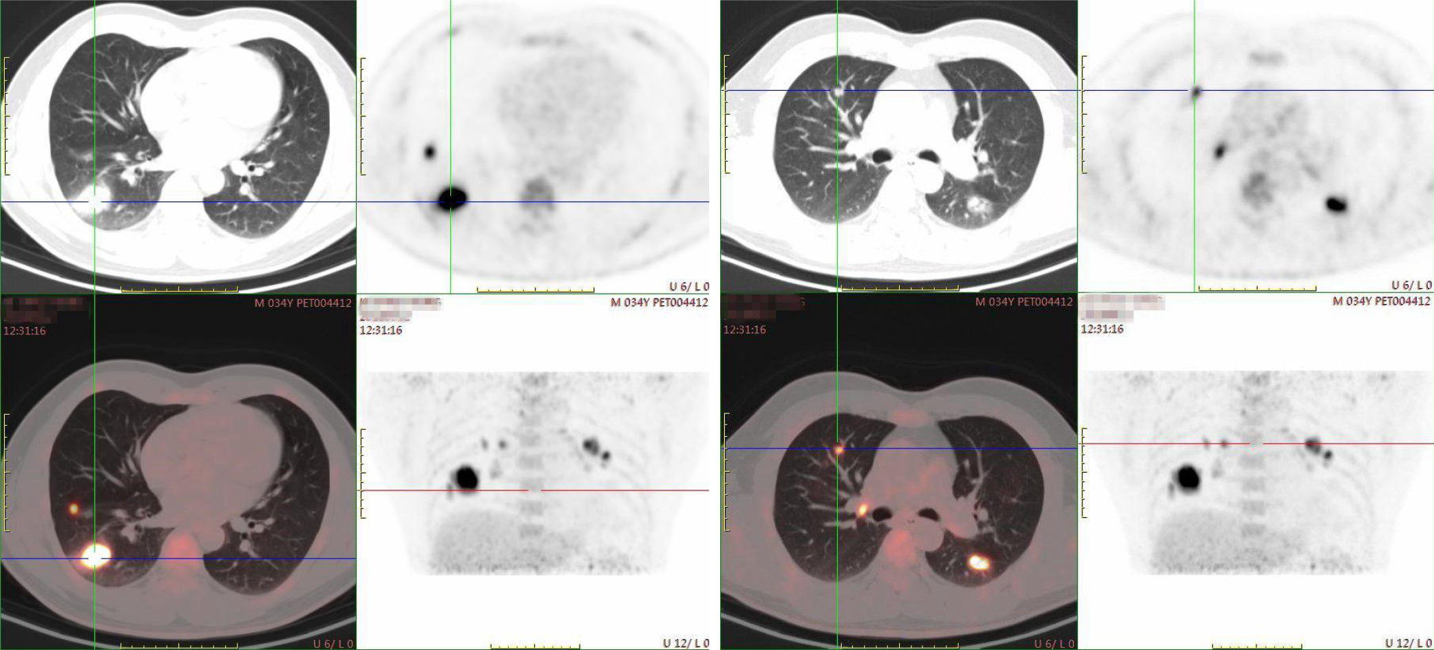
Figure 1 Positron emission tomography/computed tomography showing bilateral lesions.
An elliptic mass without clear margin was shown in the right lower lobe. The size of the mass was 3.46 cm × 2.39 cm. The enlarged right hilar lymph nodes showed a high value of SUVmax. There were also scattered nodules found in both lungs.
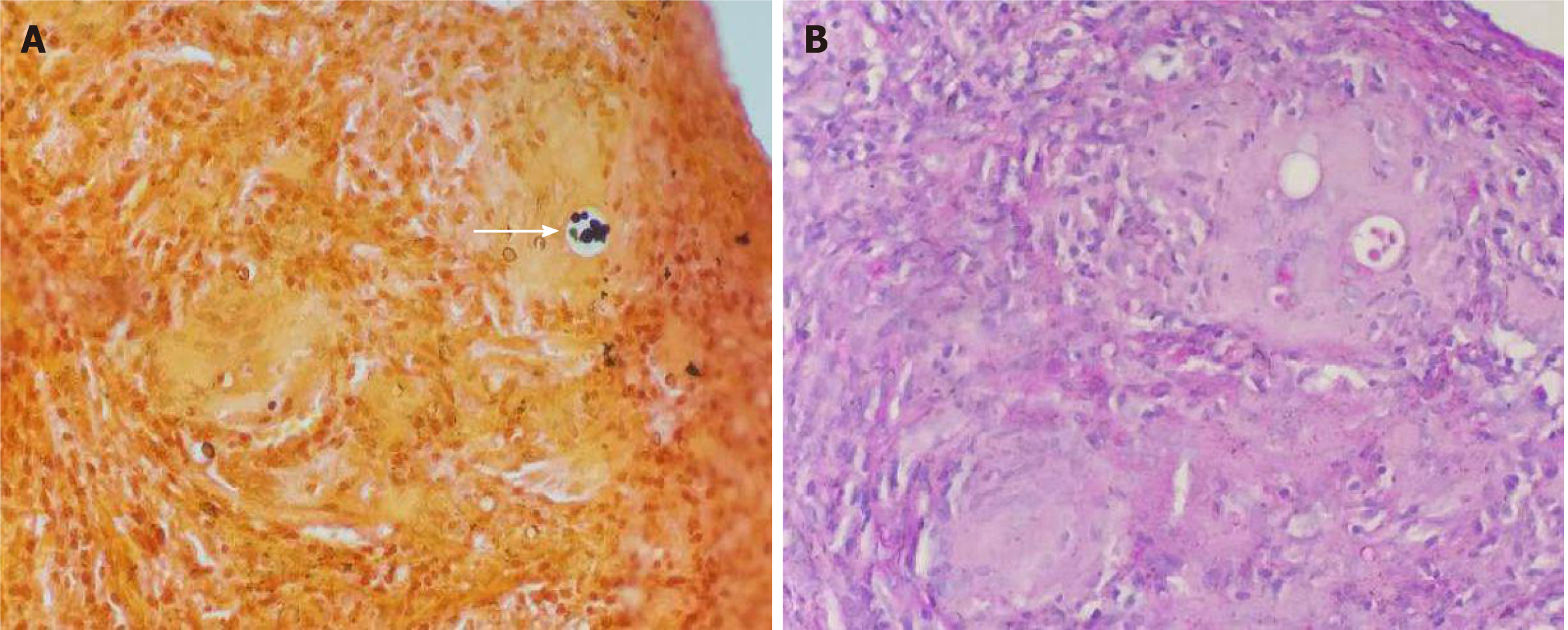
Figure 2 Pathological examination revealed Cryptococcus infection.
A: Grocott’s methenamine silver staining showed Cryptococcus spores by black staining (arrow); B: Periodic acid-Schiff staining was negative. Original magnification: × 400.
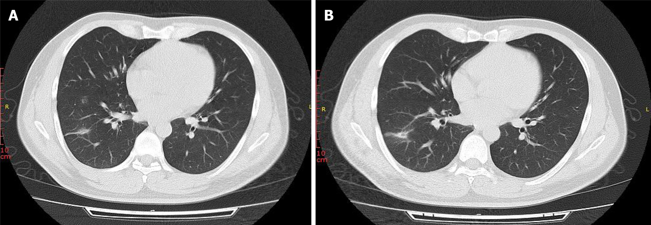
Figure 3 Chest computed tomography scan after 1 mo of antifungal treatment showing resolution of the bilateral lesions.
The mass in the right lung was reduced markedly. A: The 25th floor scan; B: The 26th floor scan.
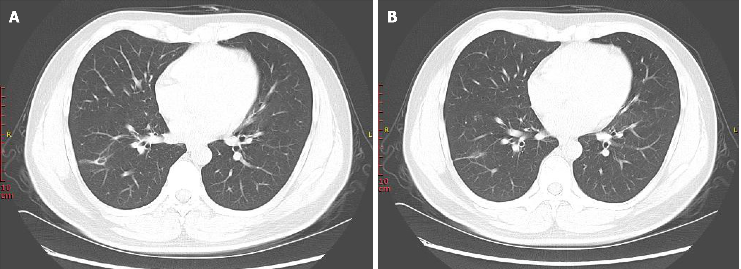
Figure 4 Chest computed tomography scan after 3 mo of antifungal treatment showing near complete disappearance of the nodules and infiltration distributed around the lesions.
Only a small number of pulmonary cavities remained at this time. A: The 29th floor scan; B: The 30th floor scan.
- Citation: Li Y, Fang L, Chang FQ, Xu FZ, Zhang YB. Cryptococcus infection with asymptomatic diffuse pulmonary disease in an immunocompetent patient: A case report. World J Clin Cases 2021; 9(11): 2619-2626
- URL: https://www.wjgnet.com/2307-8960/full/v9/i11/2619.htm
- DOI: https://dx.doi.org/10.12998/wjcc.v9.i11.2619


