Copyright
©The Author(s) 2021.
World J Clin Cases. Apr 16, 2021; 9(11): 2602-2610
Published online Apr 16, 2021. doi: 10.12998/wjcc.v9.i11.2602
Published online Apr 16, 2021. doi: 10.12998/wjcc.v9.i11.2602
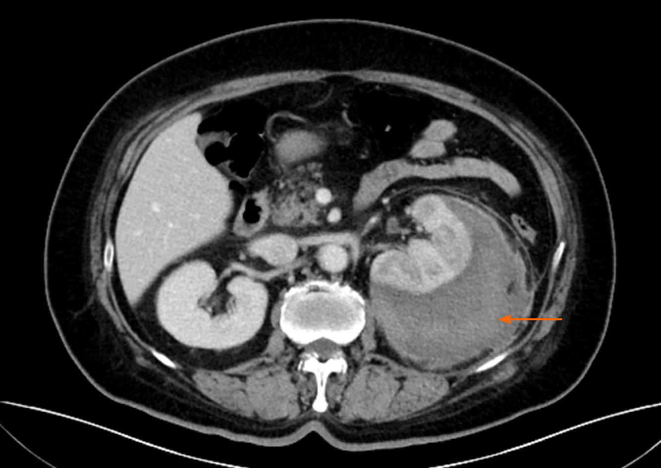
Figure 1 Venous phase of computed tomography scan of the patient.
The image shows low-density liquid dark areas of the left renal capsule.
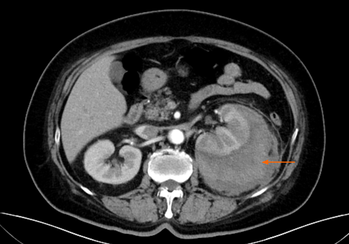
Figure 2 Arterial phase of computed tomography scan of the patient.
The image shows no enhancement of the hematoma.
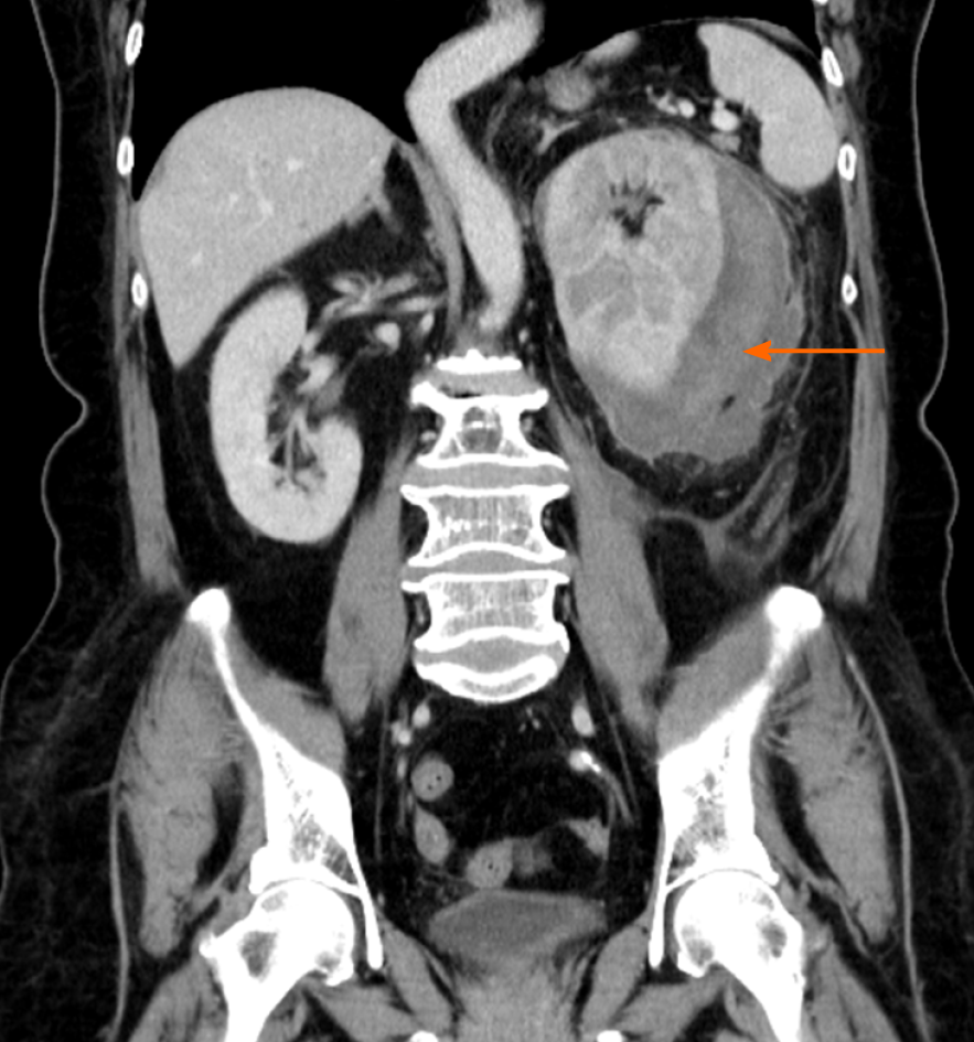
Figure 3 Contrast-enhanced computed tomography scan of the patient at the initial visit.
The left renal capsule had a crescent-shaped, low-density shadow, and the computed tomography value of the contrast-enhanced scan without enhancement was 53 HU.
- Citation: Zhang CG, Duan M, Zhang XY, Wang Y, Wu S, Feng LL, Song LL, Chen XY. Klebsiella pneumoniae infection secondary to spontaneous renal rupture that presents only as fever: A case report. World J Clin Cases 2021; 9(11): 2602-2610
- URL: https://www.wjgnet.com/2307-8960/full/v9/i11/2602.htm
- DOI: https://dx.doi.org/10.12998/wjcc.v9.i11.2602


