Copyright
©The Author(s) 2021.
World J Clin Cases. Apr 16, 2021; 9(11): 2595-2601
Published online Apr 16, 2021. doi: 10.12998/wjcc.v9.i11.2595
Published online Apr 16, 2021. doi: 10.12998/wjcc.v9.i11.2595
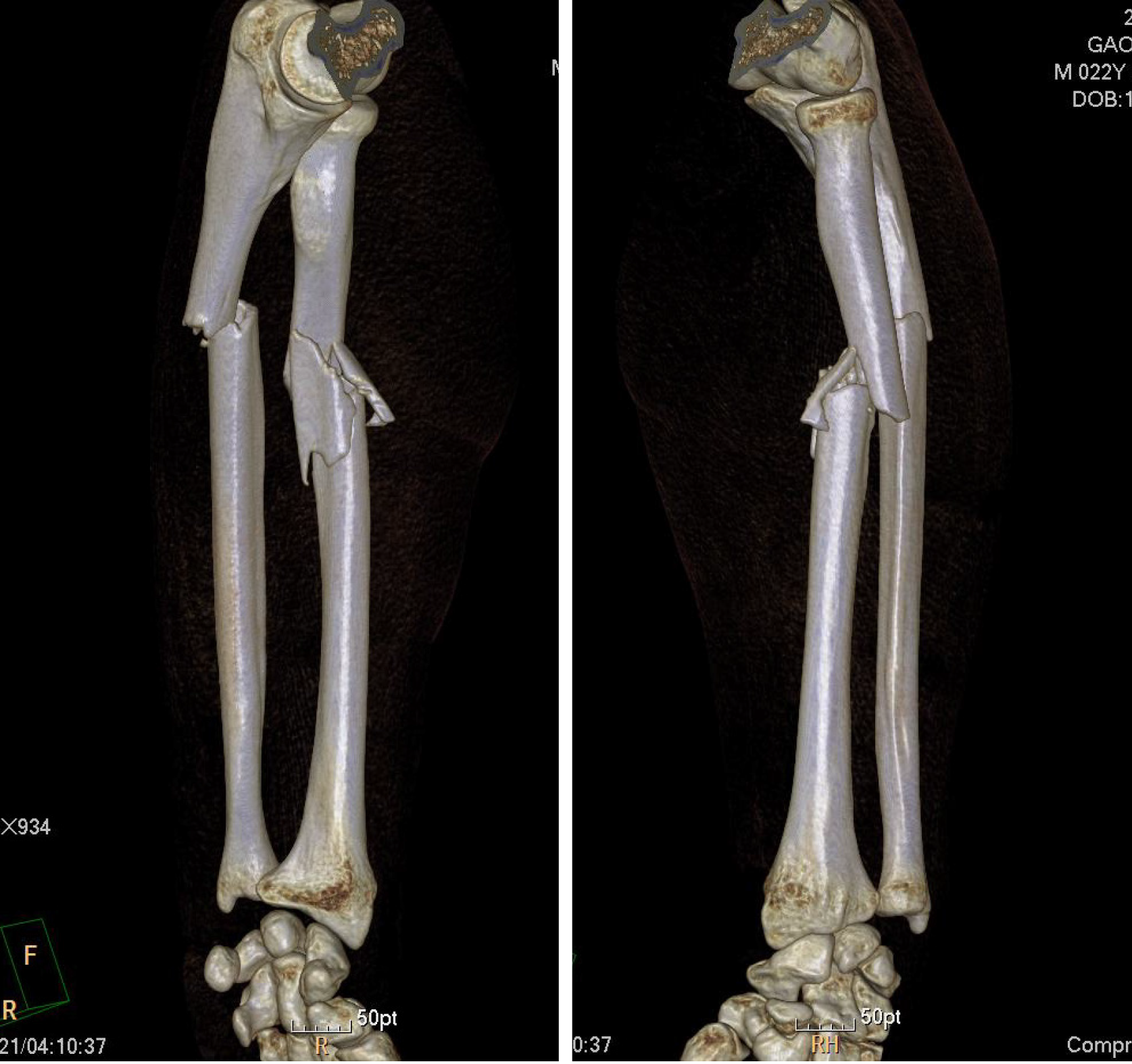
Figure 1 Three-dimensional computed tomography before surgery.
Three-dimensional computed tomography examination shows an ulna fracture of the left forearm and comminuted fracture of the radius. The fracture was located in upper third of the radius with significant displacement on the fracture side.
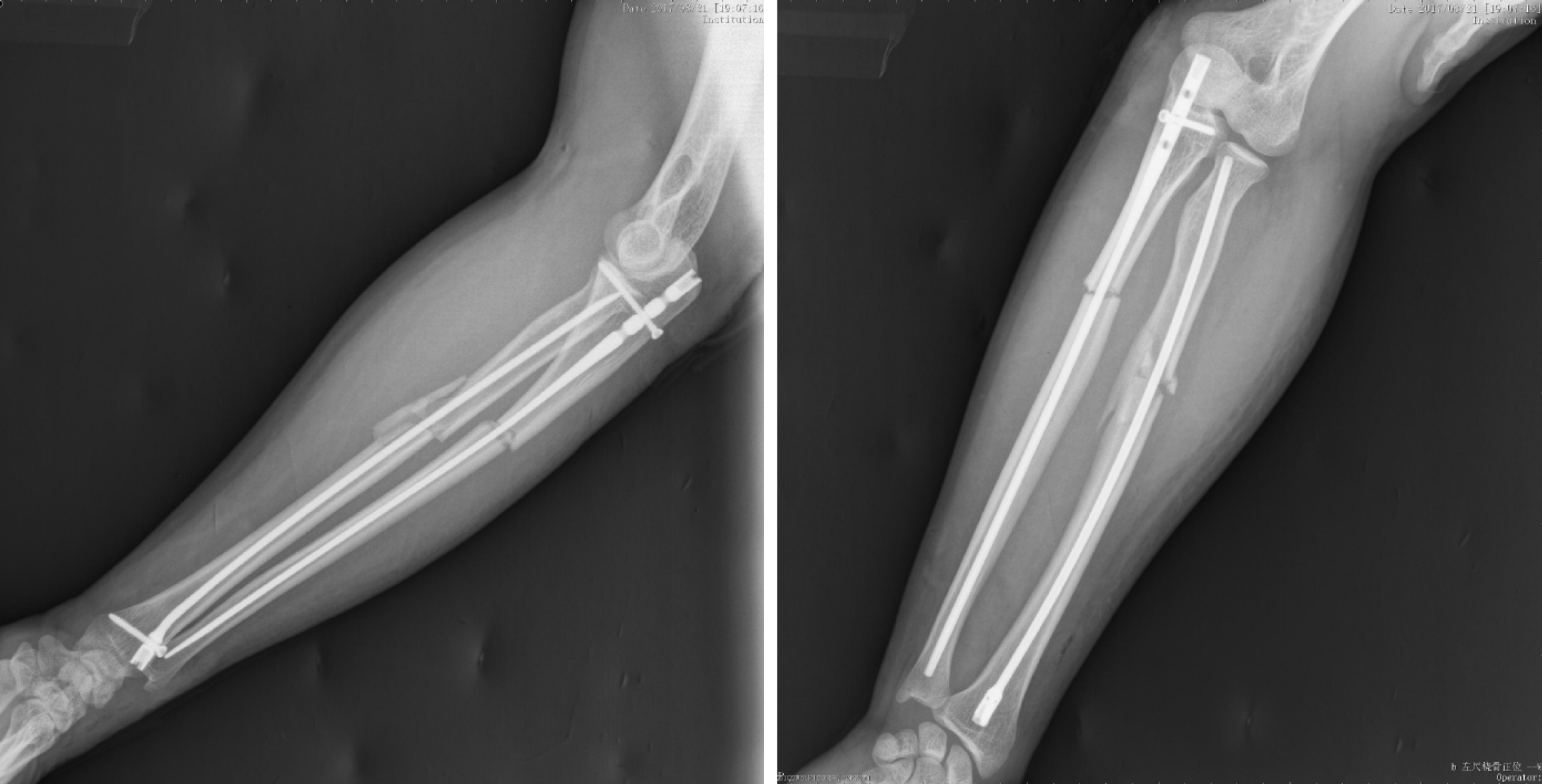
Figure 2 Imaging of the patient after surgery.
X-ray examination after surgery shows good positioning.
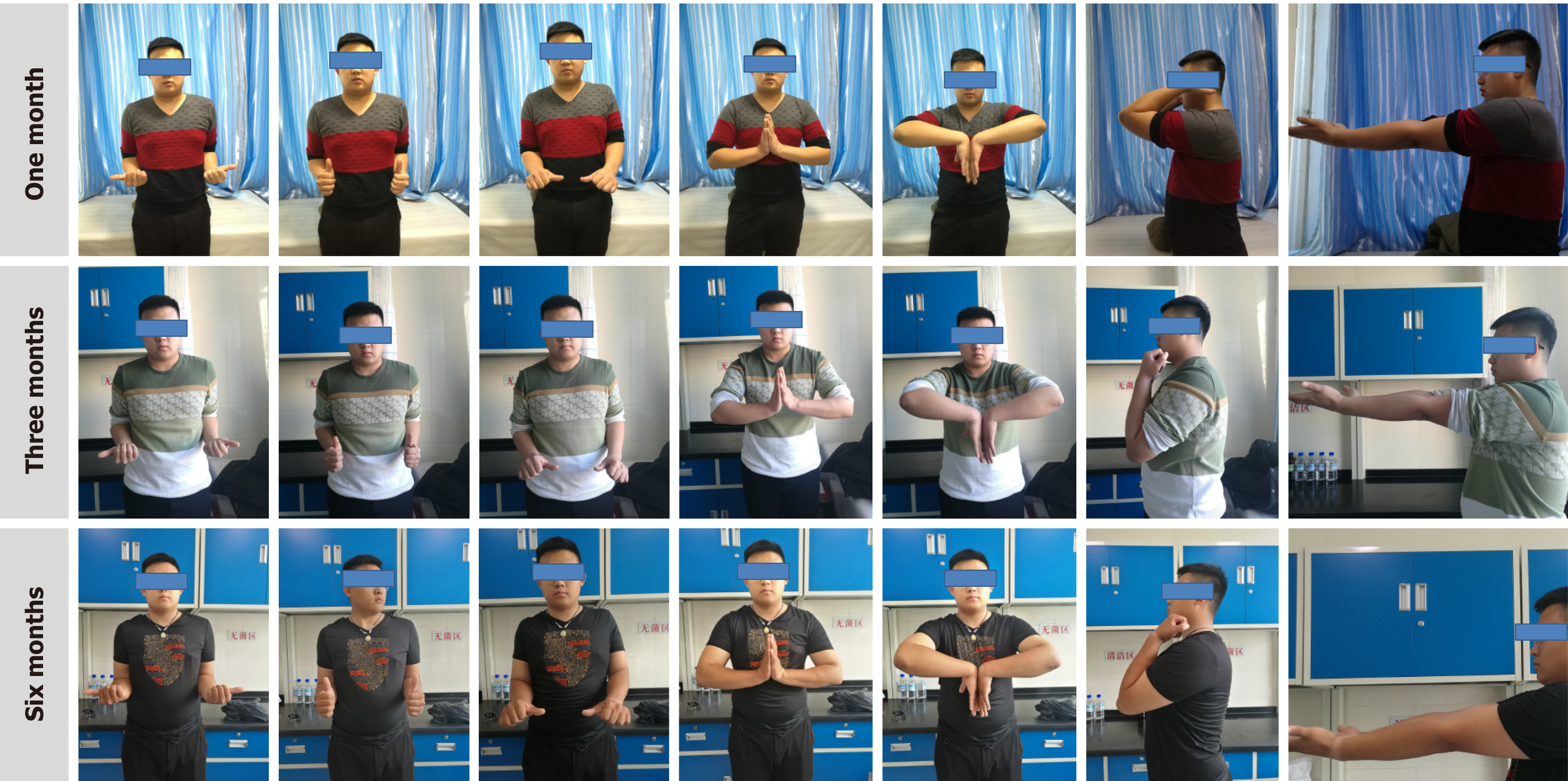
Figure 3 Follow-up photos at 1 mo, 3 mo and 6 mo after surgery.
Limb function was evaluated at 1, 3 and 6 mo after surgery. The function of the fractured limb was evaluated using the disabilities of the arm, shoulder and hand (DASH) score on a scale of 0-100, with 0 indicating that upper limb function was completely normal and 100 indicating that upper limb function was extremely limited. The DASH scores were 20 points at 1 mo, 14.2 points at 3 mo, 5 points at 6 mo, and 1.7 points at 12 mo.
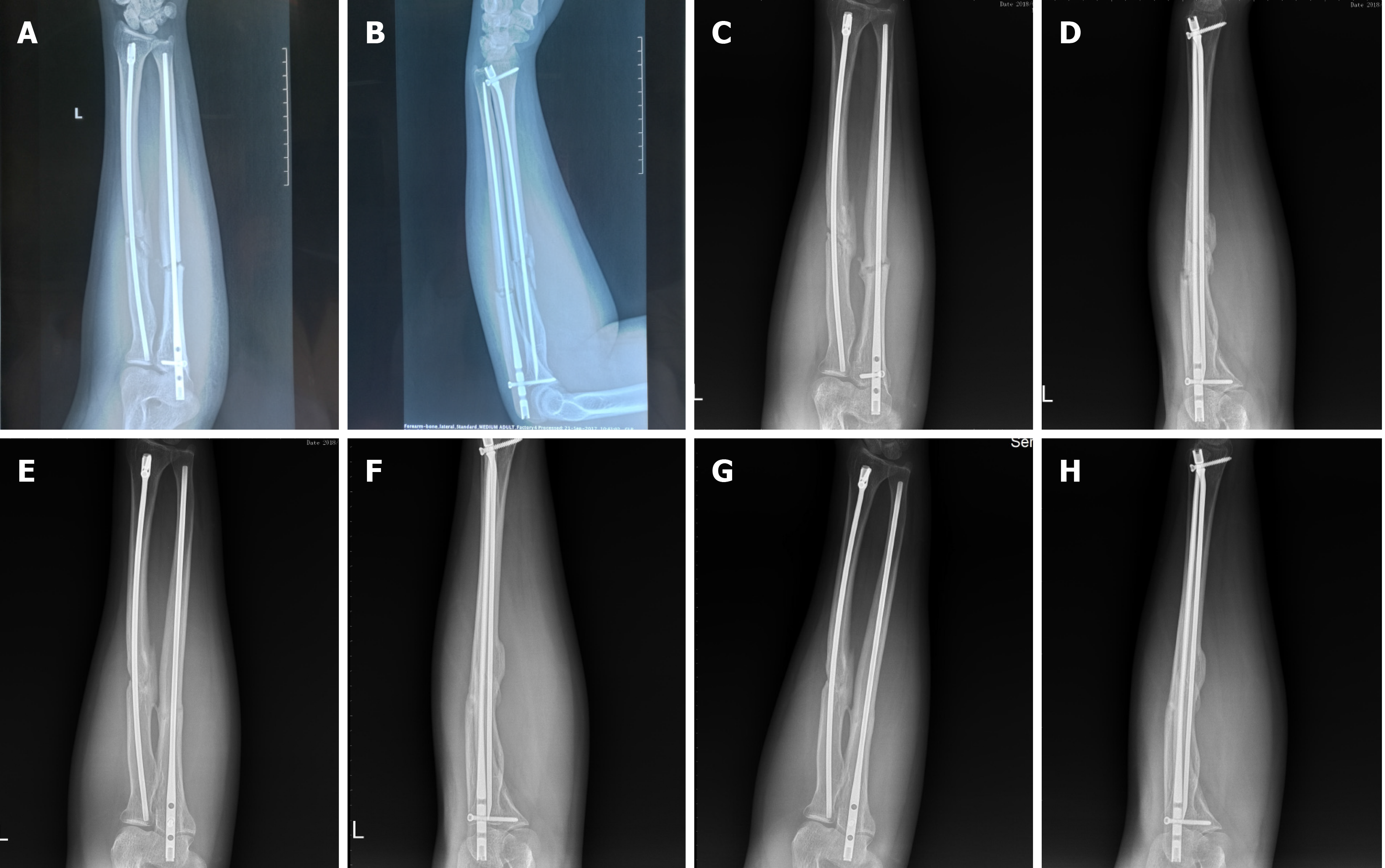
Figure 4 Imaging during follow-up.
Imaging shows complete fracture healing. No fracture line was observed. A and B: One month after the operation; C and D: Three months after the operation; E and F: Six months after the operation; G and H: Twelve months after the operation.
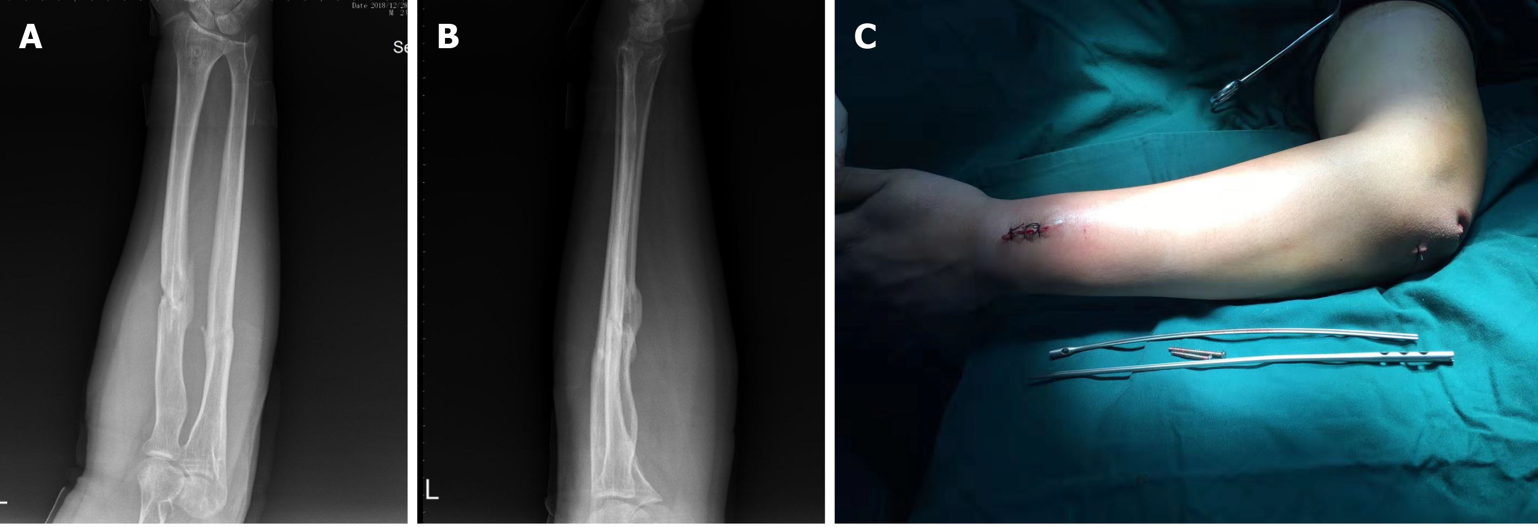
Figure 5 Imaging results after removal of internal fixation.
A and B: After successful surgery to remove the implant, the images show complete fracture healing. No fracture line was observed; C: Removal of internal fixation was completely successful.
- Citation: Liu JC, Huang BZ, Ding J, Mu XJ, Li YL, Piao CD. Minimally invasive treatment of forearm double fracture in adult using Acumed forearm intramedullary nail: A case report. World J Clin Cases 2021; 9(11): 2595-2601
- URL: https://www.wjgnet.com/2307-8960/full/v9/i11/2595.htm
- DOI: https://dx.doi.org/10.12998/wjcc.v9.i11.2595


