Copyright
©The Author(s) 2021.
World J Clin Cases. Apr 16, 2021; 9(11): 2562-2568
Published online Apr 16, 2021. doi: 10.12998/wjcc.v9.i11.2562
Published online Apr 16, 2021. doi: 10.12998/wjcc.v9.i11.2562
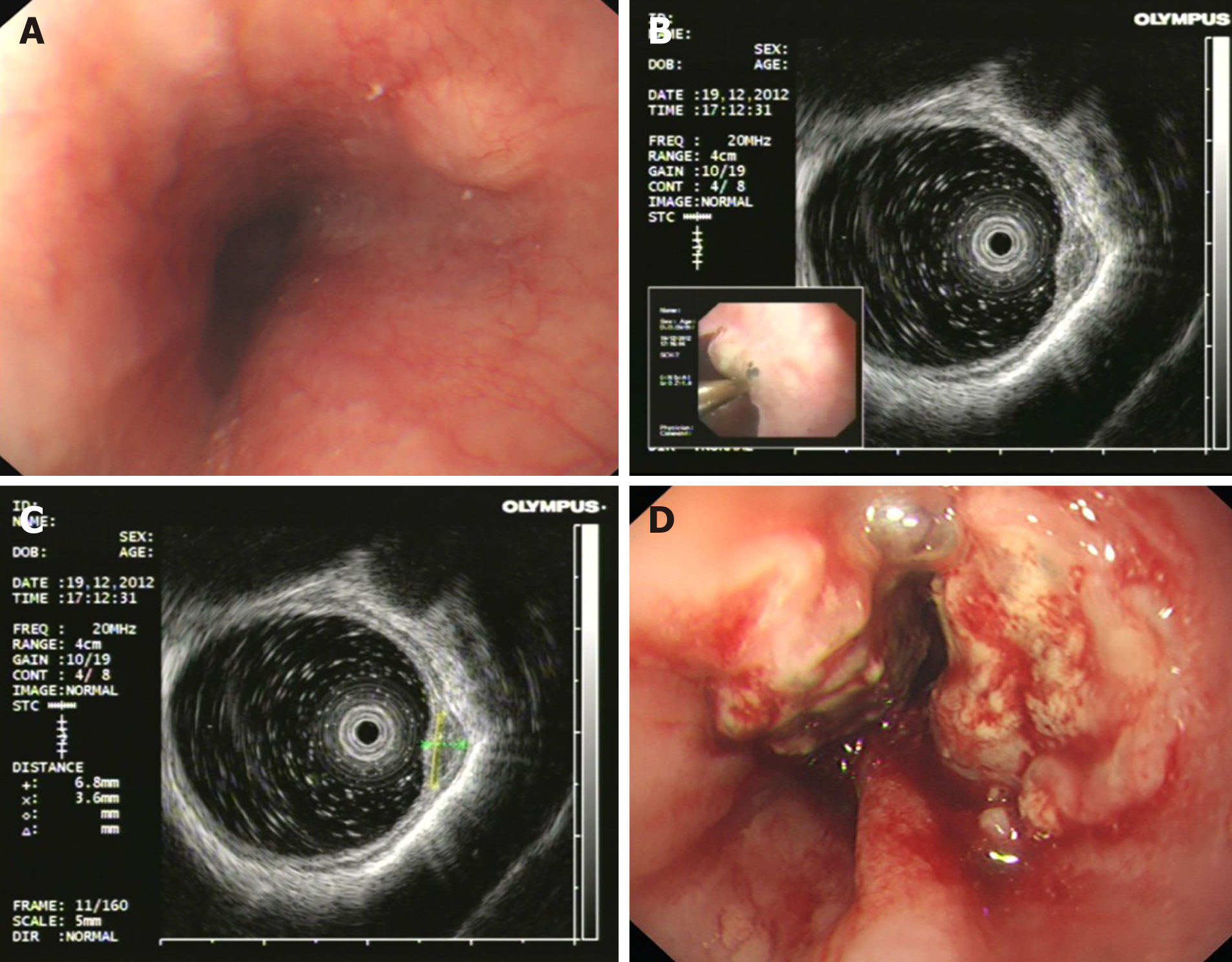
Figure 1 Findings of endoscopic examination (Case 1).
A: Esophageal submucosal tumors diagnosed by endoscopy; B: Endoscopic ultrasonography (20 MHz) showed a hypoechoic mass in the muscularis mucosa; C: The mass was about 6.8 mm × 3.6 mm in size; D: Neoplasia was detected by endoscopy 1 year later.
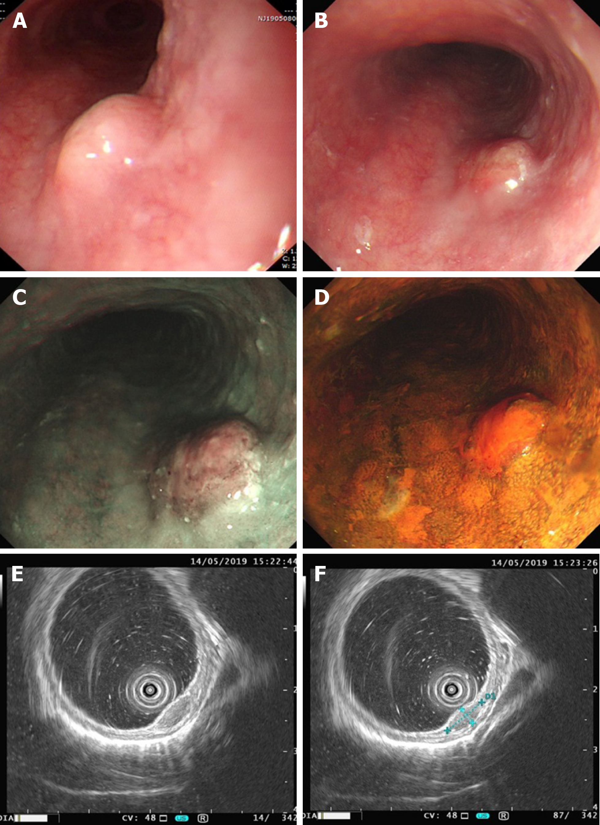
Figure 2 Findings of endoscopic examination (Case 2).
A: An esophageal submucosal tumor diagnosed by endoscopy at a local hospital; B: Mucosal hyperemia and erosion on the surface of the tumor about 2 wk later; C: Narrowband imaging showed that a portion of the depressed area was slightly brownish; D: The hyperemic and erosive mucosa of the tumor was unstained after iodine staining; E: Endoscopic ultrasonography (20 MHz) showed a hypoechoic mass in the muscularis mucosa; F: The tumor was about 7.4 mm × 2.9 mm in size.
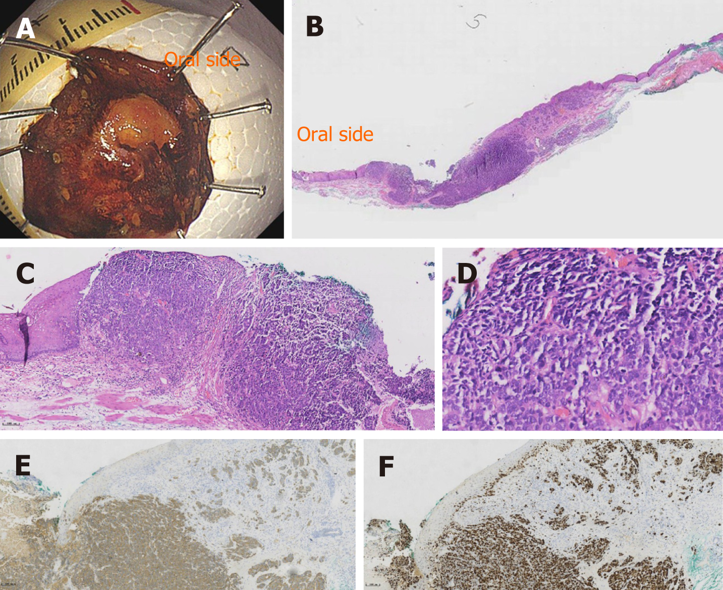
Figure 3 Histological findings of the resected specimen (Case 2).
A: The specimen resected by endoscopic submucosal dissection; B: The macroscopic appearance of the small cell carcinoma; C: The tumor was partially covered by a non-neoplastic epithelium (hematoxylin and eosin staining, magnification × 100); D: The small cell carcinoma component observed in a high power field (magnification × 400); E: Immunohistochemical staining showed that synaptophysin were positive (hematoxylin and eosin staining); E: Ki-67 index was 80%.
- Citation: Er LM, Ding Y, Sun XF, Ma WQ, Yuan L, Zheng XL, An NN, Wu ML. Endoscopic diagnosis of early-stage primary esophageal small cell carcinoma: Report of two cases. World J Clin Cases 2021; 9(11): 2562-2568
- URL: https://www.wjgnet.com/2307-8960/full/v9/i11/2562.htm
- DOI: https://dx.doi.org/10.12998/wjcc.v9.i11.2562


