Copyright
©The Author(s) 2021.
World J Clin Cases. Apr 16, 2021; 9(11): 2533-2541
Published online Apr 16, 2021. doi: 10.12998/wjcc.v9.i11.2533
Published online Apr 16, 2021. doi: 10.12998/wjcc.v9.i11.2533
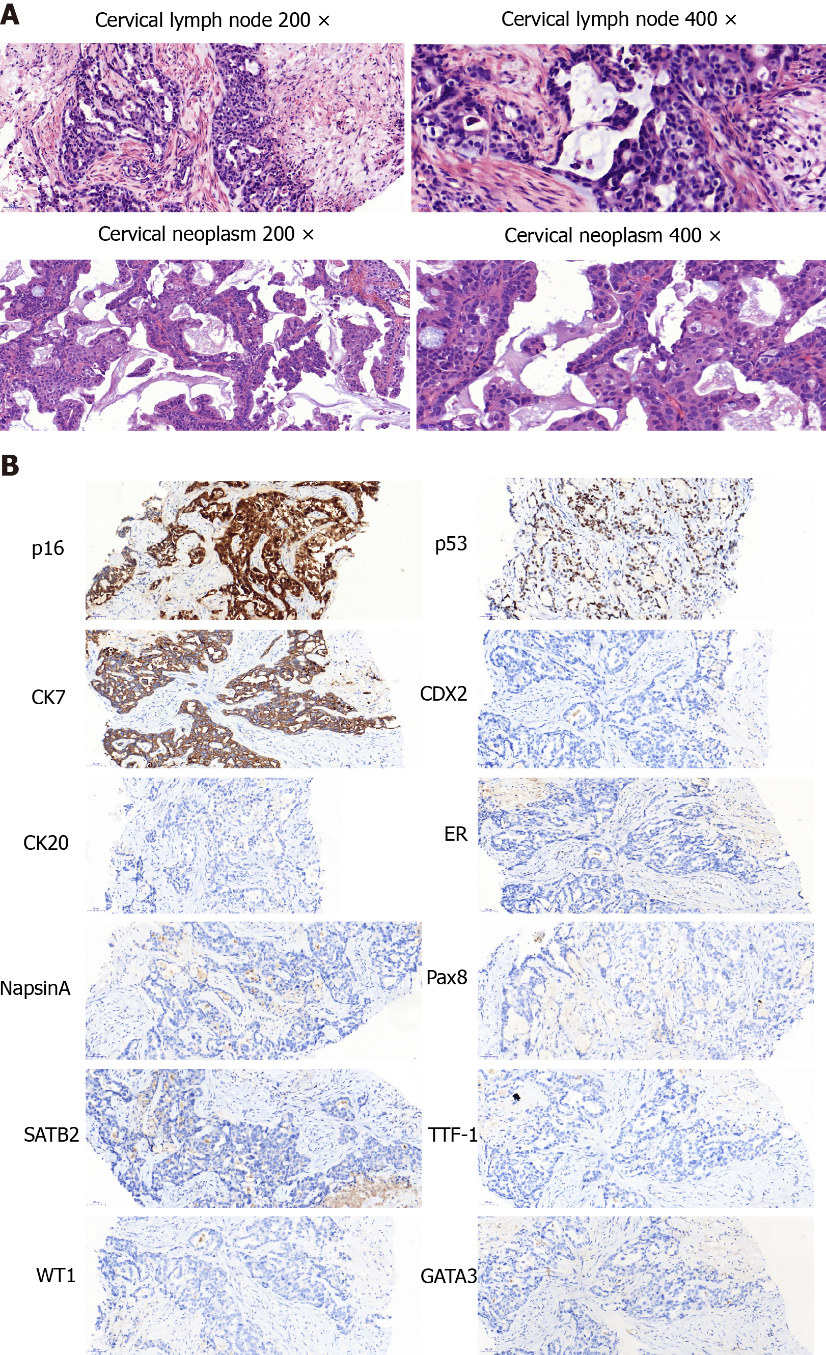
Figure 1 Pathological examination.
A and B: Cervical lymph node (A) and cervical neoplasm (B).
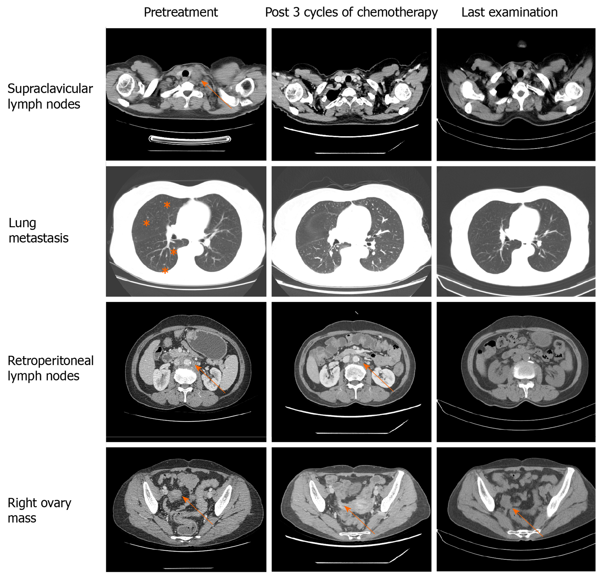
Figure 2 Computed tomography, axial view.
Arrows indicate metastatic lesions and the right ovary mass. Computed tomography indicated continuous shrinkage of the tumour.
- Citation: Wang Q, Niu XY, Feng H, Wu J, Gao W, Zhang ZX, Zou YW, Zhang BY, Wang HJ. Gastrointestinal-type chemotherapy prolongs survival in an atypical primary ovarian mucinous carcinoma: A case report. World J Clin Cases 2021; 9(11): 2533-2541
- URL: https://www.wjgnet.com/2307-8960/full/v9/i11/2533.htm
- DOI: https://dx.doi.org/10.12998/wjcc.v9.i11.2533


