Copyright
©The Author(s) 2021.
World J Clin Cases. Apr 16, 2021; 9(11): 2524-2532
Published online Apr 16, 2021. doi: 10.12998/wjcc.v9.i11.2524
Published online Apr 16, 2021. doi: 10.12998/wjcc.v9.i11.2524
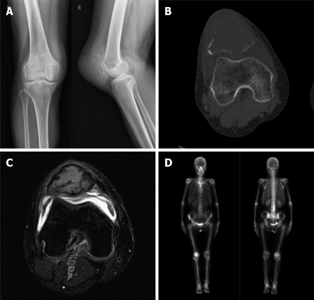
Figure 1 Preoperative radiographic results.
A: Conventional radiology, anterior posterior and lateral planes; B: Computed tomography scan, axial plane; C: Magnetic resonance imaging, axial T2; D: Bone scintigraphy.
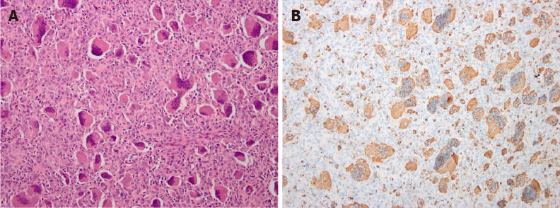
Figure 2 Pathological results.
A: Pathological specimen of the patella showed spindle-shaped mononuclear cells with osteoclastic giant cells (hematoxylin-eosin, × 200); B: Immunohistochemical expression of phosphoglucomutase 1 in tumor cells (× 200).
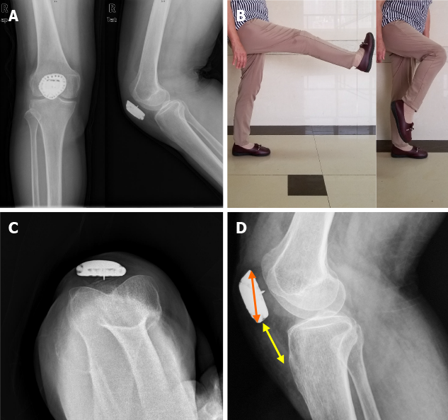
Figure 3 Postoperative radiographic results.
A: Conventional radiology, anterior posterior and lateral planes; B: Active flexion arc was 0°-120° without extension lag; C: Patellar axial radiography in maximum knee flexion (120°); D: Patella height.
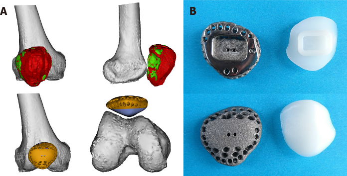
Figure 4 Design detail and prosthesis image.
A: Prosthetic design in software; B: Prosthetic profile.
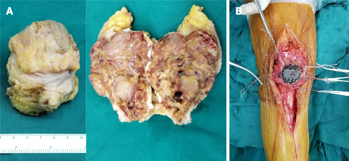
Figure 5 Specimen photos and intraoperative image.
A: Total patellectomy; B: Soft tissue reconstruction.
- Citation: Wang J, Zhou Y, Wang YT, Min L, Zhang YQ, Lu MX, Tang F, Luo Y, Zhang YH, Zhang XL, Tu CQ. Three-dimensional-printed custom-made patellar endoprosthesis for recurrent giant cell tumor of the patella: A case report and review of the literature. World J Clin Cases 2021; 9(11): 2524-2532
- URL: https://www.wjgnet.com/2307-8960/full/v9/i11/2524.htm
- DOI: https://dx.doi.org/10.12998/wjcc.v9.i11.2524


